 Login / Register


- Clone
- NVG-2 (See other available formats)
- Regulatory Status
- RUO
- Other Names
- LIMP, LAMP-3, gp55, melanoma-associated antigen, ME491
- Isotype
- Rat IgG2a, κ
- Ave. Rating
- Submit a Review
- Product Citations
- publications

-

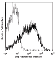
Thioglycolate-elicited BALB/c mouse peritoneal macrophages were fixed, permeabilized and intracellularly stained with purified CD63 (clone NVG-2) (left) or purified rat IgG2a, κ isotype control (right), followed by biotinylated anti Rat IgG, SAV PE, and CD11b APC. -

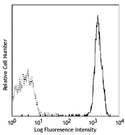
bEnd.3, mouse endothelial cells were stained with purified CD63 (clone NVG-2) (filled histogram) or rat IgG2a, κ isotype control (open histogram), followed by anti-rat IgG PE.
| Cat # | Size | Price | Quantity Check Availability | Save | ||
|---|---|---|---|---|---|---|
| 143901 | 25 µg | 72€ | ||||
| 143902 | 100 µg | 165€ | ||||
CD63, also known as LIMP, LAMP-3, gp55, and melanoma-associated antigen (ME491), is a member of the tetraspanin superfamily (TM4SF) that constitutes a main component of the lysosomal membrane. It is expressed on activated platelets, monocyte/macrophages, endothelium, fibroblasts, osteoblasts, and smooth muscle cells. CD63 may be involved in platelet activation and is thought to function as a transmembrane adaptor protein. CD63 has been shown to associate with CD9, CD81, VLA-3, and VLA-6. In mice, there are two CD63 gene loci, of which only one is functional. CD63 deficient mice are viable, and there is no alteration in the population of immune cells. A recent report shows that CD63-deficient mice exhibit a significant reduction in both leukocyte rolling and recruitment in a peritonitis model.
Product DetailsProduct Details
- Verified Reactivity
- Mouse
- Antibody Type
- Monoclonal
- Host Species
- Rat
- Immunogen
- Intestinal lamina propria light-density cells (enriched with eosinophils)
- Formulation
- Phosphate-buffered solution, pH 7.2, containing 0.09% sodium azide.
- Preparation
- The antibody was purified by affinity chromatography.
- Concentration
- 0.5 mg/mL
- Storage & Handling
- The antibody solution should be stored undiluted between 2°C and 8°C.
- Application
-
ICFC - Quality tested
FC - Verified
WB, IHC-F - Reported in the literature, not verified in house - Recommended Usage
-
Each lot of this antibody is quality control tested by intracellular immunofluorescent staining with flow cytometric analysis. For intracellular flow cytometric staining, the suggested use of this reagent is ≤ 0.5 µg per million cells in 100 µL volume. For flow cytometric staining, the suggested use of this reagent is ≤ 1.0 µg per million cells in 100 µL volume. It is recommended that the reagent be titrated for optimal performance for each application.
- Application Notes
-
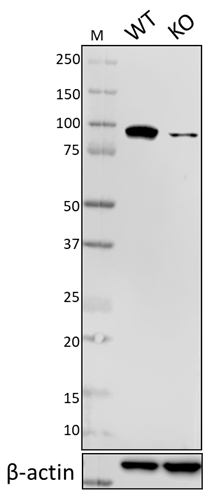
Additional reported applications (for the relevant formats) include: Western blotting1 and immunofluorescence1.
-
Application References
(PubMed link indicates BioLegend citation) -
- Verjan Garcia N, et al. 2011. J. Immunol. 187:2268. (WB, IF)
- Product Citations
-
- RRID
-
AB_11203908 (BioLegend Cat. No. 143901)
AB_11204263 (BioLegend Cat. No. 143902)
Antigen Details
- Structure
- Tetraspan transmembrane superfamily (TM4SF), type III lysosomal glycoprotein, 53 kD
- Distribution
-
Activated platelets, monocytes, macrophages, endothelium, fibroblasts, osteoclasts, and smooth muscle cells
- Function
- Platelet activation
- Interaction
- CD9, CD81, VLA-3, and VLA-6
- Cell Type
- Endothelial cells, Fibroblasts, Macrophages, Monocytes, Osteoclasts, Platelets
- Biology Area
- Immunology, Innate Immunity
- Molecular Family
- CD Molecules
- Antigen References
-
1. Azorsa DO, et al. 1991. Blood 78:280.
2. Kishimoto T, et al. 1997. Leukocyte Typing V1. Oxford University Press New York.
3. Hildreth JE, et al. 1991. Blood 77:121. - Gene ID
- 12512 View all products for this Gene ID
- UniProt
- View information about CD63 on UniProt.org


Other Formats
View All CD63 Reagents Request Custom Conjugation| Description | Clone | Applications |
|---|---|---|
| Purified anti-mouse CD63 | NVG-2 | ICFC,FC,WB,IHC-F |
| PE anti-mouse CD63 | NVG-2 | ICFC,FC |
| APC anti-mouse CD63 | NVG-2 | ICFC,FC |
| APC/Cyanine7 anti-mouse CD63 | NVG-2 | ICFC,FC |
| PE/Dazzle™ 594 anti-mouse CD63 | NVG-2 | ICFC,FC |
| PE/Cyanine7 anti-mouse CD63 | NVG-2 | ICFC,FC |
| PerCP/Cyanine5.5 anti-mouse CD63 | NVG-2 | ICFC,FC |
| TotalSeq™-A0559 anti-mouse CD63 | NVG-2 | PG |
| FITC anti-mouse CD63 | NVG-2 | ICFC,FC |
| Biotin anti-mouse CD63 | NVG-2 | ICFC,FC |
| Alexa Fluor® 647 anti-mouse CD63 | NVG-2 | ICFC,FC |
| Alexa Fluor® 700 anti-mouse CD63 | NVG-2 | ICFC,FC |
| TotalSeq™-C0559 anti-mouse CD63 | NVG-2 | PG |
| TotalSeq™-B0559 anti-mouse CD63 | NVG-2 | PG |
Customers Also Purchased
Compare Data Across All Formats
This data display is provided for general comparisons between formats.
Your actual data may vary due to variations in samples, target cells, instruments and their settings, staining conditions, and other factors.
If you need assistance with selecting the best format contact our expert technical support team.
-
Purified anti-mouse CD63

Thioglycolate-elicited BALB/c mouse peritoneal macrophages w... 
bEnd.3, mouse endothelial cells were stained with purified C... -
PE anti-mouse CD63

Thioglycolate-elicited BALB/c mouse peritoneal macrophages w... 
bEnd.3, mouse endothelial cells were stained with CD63 (clon... -
APC anti-mouse CD63

Thioglycolate-elicited BALB/c mouse peritoneal macrophages w... 
bEND.3 cells were stained with CD63 (clone NVG-2) APC (fille... -
APC/Cyanine7 anti-mouse CD63

C57BL/6 resident peritoneal cells were stained with CD11b FI... 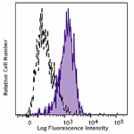
-
PE/Dazzle™ 594 anti-mouse CD63

Thioglycolate-elicited BALB/c mouse peritoneal macrophages w... 
bEND.3 cells were stained with CD63 (clone NVG-2) PE/ Dazzle... -
PE/Cyanine7 anti-mouse CD63

Thioglycolate-elicited BALB/c mouse peritoneal macrophages w... 
bEND.3 cells were stained with CD63 (clone NVG-2) PE/Cyanine... -
PerCP/Cyanine5.5 anti-mouse CD63

glycolate-elicited BALB/c mouse peritoneal macrophages were ... 
bEND.3 cells were stained with CD63 (clone NVG-2) PerCp/Cy5.... -
TotalSeq™-A0559 anti-mouse CD63
-
FITC anti-mouse CD63

Thioglycolate-elicited BALB/c mouse peritoneal macrophages w... 
Bend.3 mouse endothelial cells were stained with CD63 (clone... -
Biotin anti-mouse CD63

Thioglycollate-elicited BALB/c mouse peritoneal macrophages ... 
Bend.3 mouse endothelial cells were stained with biotinylate... -
Alexa Fluor® 647 anti-mouse CD63

Thioglycolate-elicited BALB/c mouse peritoneal macrophages w... 
bEnd.3, mouse endothelial cells were stained with CD63 (clon... -
Alexa Fluor® 700 anti-mouse CD63

Thioglycolate-elicited BALB/c mouse peritoneal macrophages w... 
bEnd.3, mouse endothelial cells were stained with CD63 (clon... -
TotalSeq™-C0559 anti-mouse CD63
-
TotalSeq™-B0559 anti-mouse CD63


Follow Us