 Login / Register


- Clone
- 2H7 (See other available formats)
- Regulatory Status
- RUO
- Workshop
- IV B201
- Other Names
- B1, Bp35
- Isotype
- Mouse IgG2b, κ
- Ave. Rating
- Submit a Review
- Product Citations
- publications

-

Human frozen tonsil section was fixed with 4% paraformaldehyde (PFA) for ten minutes and blocked with 5% FBS for 30 minutes at room temperature. Then the section was stained with 10 µg/ml purified anti-human CD20 (clone 2H7) overnight at 4°C, followed by 2.5 µg/ml of anti-mouse IgG (clone Poly4053) Alexa Fluor® 594 (red) for two hours at room temperature. Nuclei were counterstained with DAPI (blue). The image was captured by 10X objective. -

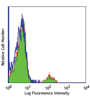
Human peripheral blood lymphocytes were stained with purified anti-CD20 (clone 2H7) (solid line), or mouse IgG2b, κ (dashed line) followed by anti-mouse IgGs FITC.
| Cat # | Size | Price | Quantity Check Availability | Save | ||
|---|---|---|---|---|---|---|
| 302301 | 25 µg | 34€ | ||||
| 302302 | 100 µg | 63€ | ||||
CD20 is a 33-37 kD, four transmembrane spanning protein, also known as B1 and Bp35. CD20 is expressed on pre-B-cells, resting and activated B cells (not plasma cells), some follicular dendritic cells, and at low levels on a T cell subset. CD20 is heavily phosphorylated on activated B cells and malignant B cells. Homo-oligomeric complexes of CD20 are thought to form Ca2+ conductive ion channels in the plasma membrane of B cells. The CD20 molecule is involved in B-cell activation and is associated with various Src family kinases (Lyn, Lck, Fyn). It exists in a complex with MHC class I and II, CD53, CD81, and CD82.
Product DetailsProduct Details
- Verified Reactivity
- Human, Cynomolgus, Rhesus
- Reported Reactivity
- Baboon, Capuchin Monkey, Chimpanzee, Pigtailed Macaque, Squirrel Monkey
- Antibody Type
- Monoclonal
- Host Species
- Mouse
- Immunogen
- Human tonsillar B cells
- Formulation
- Phosphate-buffered solution, pH 7.2, containing 0.09% sodium azide.
- Preparation
- The antibody was purified by affinity chromatography.
- Concentration
- 0.5 mg/ml
- Storage & Handling
- The antibody solution should be stored undiluted between 2°C and 8°C.
- Application
-
FC - Quality tested
CyTOF®, IHC-F - Verified
IP - Reported in the literature, not verified in house - Recommended Usage
-
Each lot of this antibody is quality control tested by immunofluorescent staining with flow cytometric analysis. For flow cytometric staining, the suggested use of this reagent is ≤2.0 µg per million cells in 100 µl volume. For immunohistochemical staining on frozen tissue sections, the suggested use of this reagent is 5.0 - 10 µg per ml. It is recommended that the reagent be titrated for optimal performance for each application.
- Application Notes
-
The epitope recognized by clone 2H7 has been mapped to the sequence YNCEPANPSEKNSPST which lies in the large extracellular loop of human CD20. Additional reported applications (for the relevant formats) include: immunoprecipitation4 and immunohistochemical staining of acetone-fixed frozen sections5.
-
Application References
(PubMed link indicates BioLegend citation) -
- Schlossman S, et al. 1995. Leucocyte Typing V. Oxford University Press. New York.
- Knapp W, et al. 1989. Leucocyte Typing IV. Oxford University Press. New York.
- McMichael A, et al. Eds. 1987. Leucocyte Typing III Oxford University Press. New York.
- Polyak MJ, et al. 2002. Blood 99:3256. (IP)
- Mack CL, et al. 2004. Pediatr. Res. 56:79. (IHC)
- Product Citations
-
- RRID
-
AB_314249 (BioLegend Cat. No. 302301)
AB_314250 (BioLegend Cat. No. 302302)
Antigen Details
- Structure
- Four transmembrane protein (TM4SF), heavily phosphorylated after activation, 33-37 kD
- Distribution
-
B cell, T cell subsets
- Function
- B cell activation
- Ligand/Receptor
- Src family tyrosine kinases, MHC class I, II, CD53, CD81, CD82
- Cell Type
- B cells, T cells
- Biology Area
- Costimulatory Molecules, Immunology
- Molecular Family
- CD Molecules
- Antigen References
-
1. Hultin L, et al. 1993. Cytometry 14:196.
2. Tedder T, et al. 1994. Immunol. Today 15:450. - Gene ID
- 931 View all products for this Gene ID
- UniProt
- View information about CD20 on UniProt.org


Related FAQs
- What is the difference between two anti human CD20 clones 2H7 and 1412?
-
For clone 1412: This clone specifically recognizes cytoplasmic domain of CD20 and thus can only be used for intracellular flow cytometry. In this instance you will need to include the fixation and permeabilization steps. Please follow the intracellular flow cytometry staining protocol.
For clone 2H7: This clone is ok for regular surface staining for CD20 and there is no need for any fixation and permeabilization steps.
Our technical protocols can be found here.
Other Formats
View All CD20 Reagents Request Custom ConjugationCustomers Also Purchased
Compare Data Across All Formats
This data display is provided for general comparisons between formats.
Your actual data may vary due to variations in samples, target cells, instruments and their settings, staining conditions, and other factors.
If you need assistance with selecting the best format contact our expert technical support team.
-
APC anti-human CD20

Human peripheral blood lymphocytes were stained with anti-CD... -
FITC anti-human CD20

Human peripheral blood lymphocytes were stained with anti-CD... -
PE anti-human CD20

Human peripheral blood lymphocytes were stained with anti-CD... -
PE/Cyanine5 anti-human CD20

Human peripheral blood lymphocytes were stained with anti-CD... -
Purified anti-human CD20

Human peripheral blood lymphocytes were stained with purifie... 
Human frozen tonsil section was fixed with 4% paraformaldehy... -
APC/Cyanine7 anti-human CD20

Human peripheral blood lymphocytes were stained with anti-CD... -
PE/Cyanine7 anti-human CD20

Human peripheral blood lymphocytes were stained with anti-CD... -
Alexa Fluor® 488 anti-human CD20

Human peripheral blood lymphocytes were stained with anti-CD... 
Human frozen tonsil section was fixed with 4% paraformaldehy... -
Alexa Fluor® 647 anti-human CD20

Human peripheral blood lymphocytes were stained with anti-CD... 
Human frozen tonsil section was fixed with 4% paraformaldehy... -
Pacific Blue™ anti-human CD20

Human peripheral blood lymphocytes were stained with anti-CD... -
Alexa Fluor® 700 anti-human CD20

Human peripheral blood lymphocytes were stained with anti-CD... -
PerCP anti-human CD20

Human peripheral blood lymphocytes were stained with anti-CD... -
PerCP/Cyanine5.5 anti-human CD20

Human peripheral blood lymphocytes were stained with CD19 AP... -
Brilliant Violet 421™ anti-human CD20

Human peripheral blood lymphocytes were stained with CD19 PE... 

Human frozen tonsil section was fixed with 4% paraformaldehy... -
Brilliant Violet 570™ anti-human CD20

Human peripheral blood lymphocytes were stained with CD20 (c... -
Brilliant Violet 605™ anti-human CD20

Human peripheral blood lymphocytes were stained with CD20 (c... -
Brilliant Violet 650™ anti-human CD20

Human peripheral blood lymphocytes were stained with CD20 (c... -
Brilliant Violet 785™ anti-human CD20

Human peripheral blood lymphocytes were stained with CD20 (c... -
Brilliant Violet 510™ anti-human CD20

Human peripheral blood lymphocytes were stained with CD20 (c... -
Brilliant Violet 711™ anti-human CD20

Human peripheral blood lymphocytes were stained with CD20 (c... -
Purified anti-human CD20 (Maxpar® Ready)

Human PBMCs stained with 154Sm-anti-CD45 (HI30) and 147m-ant... -
PE/Dazzle™ 594 anti-human CD20

Human peripheral blood lymphocytes were stained with CD20 PE... -
Biotin anti-human CD20

Human peripheral blood lymphocytes were stained with biotiny... -
APC/Fire™ 750 anti-human CD20

Human peripheral blood lymphocytes were stained with CD3 PE ... 
-
Alexa Fluor® 594 anti-human CD20

Human peripheral blood mononuclear cells were fixed with 2% ... 
Human frozen tonsil section was fixed with 4% paraformaldehy... -
FITC anti-human CD20

Typical results from human peripheral blood lymphocytes stai... -
Pacific Blue™ anti-human CD20

Typical results from human peripheral blood lymphocytes stai... -
APC anti-human CD20

Typical results from human peripheral blood lymphocytes stai... -
PE/Cyanine7 anti-human CD20
Typical results from human peripheral blood lymphocytes stai... -
TotalSeq™-A0100 anti-human CD20
-
TotalSeq™-B0100 anti-human CD20
-
TotalSeq™-C0100 anti-human CD20
-
PerCP/Cyanine5.5 anti-human CD20

Typical results from human peripheral blood lymphocytes stai... -
Spark NIR™ 685 anti-human CD20

Human peripheral blood lymphocytes were stained with CD19 Br... -
Spark YG™ 593 anti-human CD20

Human peripheral blood lymphocytes were stained with CD19 Al... -
GMP FITC anti-human CD20

Typical results from human peripheral blood lymphocytes stai... -
TotalSeq™-D0100 anti-human CD20
-
GMP APC anti-human CD20

Typical results from human peripheral blood lymphocytes stai... -
Spark Violet™ 500 anti-human CD20

Human peripheral blood lymphocytes were stained with anti-hu... -
GMP Pacific Blue™ anti-human CD20

Typical results from human peripheral blood lymphocytes stai... -
GMP PerCP/Cyanine5.5 anti-human CD20

Typical results from human peripheral blood lymphocytes stai... -
Spark Violet™ 538 anti-human CD20

Human peripheral blood lymphocytes were stained with anti-hu... -
APC/Fire™ 750 anti-human CD20

Typical results from human peripheral blood lymphocytes stai... -
PE anti-human CD20

Typical results from Human peripheral blood lymphocytes stai... -
GMP PE/Cyanine7 anti-human CD20

Typical results from human peripheral blood lymphocytes stai... -
Spark Blue™ 515 anti-human CD20

Human peripheral blood lymphocytes were stained with anti-hu... -
GMP APC/Fire™ 750 anti-human CD20

Typical results from human peripheral blood lymphocytes stai... -
GMP PE anti-human CD20

Typical results from human peripheral blood lymphocytes stai... -
Spark Blue™ 550 anti-human CD20 (Flexi-Fluor™)
-
Spark Blue™ 574 anti-human CD20 (Flexi-Fluor™)
-
Spark Red™ 718 anti-human CD20 (Flexi-Fluor™)


Follow Us