 Login / Register


- Clone
- 1B1-B2 (See other available formats)
- Regulatory Status
- RUO
- Other Names
- Histone H3.1, histone H3/a, histone 1, H3a, H3 histone family, member A, H3/A, H3FA
- Isotype
- Mouse IgG3, κ
- Ave. Rating
- Submit a Review
- Product Citations
- publications

-

IHC staining of purified anti-Histone H3 (C-Terminus) antibody (clone 1B1-B2) on formalin-fixed paraffin-embedded rat brain tissue. Following antigen retrieval using Sodium Citrate H.I.E.R., the tissue was incubated with 1 µg/mL of the primary antibody overnight at 4°C. BioLegend's Ultra Streptavidin (USA) HRP kit (Multi-Species, DAB, Cat. No. 929901) was used for detection followed by hematoxylin counterstaining, according to the protocol provided. The image was captured with a 40X objective. -

IHC staining of purified anti-Histone H3 (C-Terminus) antibody (clone 1B1-B2) on formalin-fixed paraffin-embedded mouse brain tissue. Following antigen retrieval using Sodium Citrate H.I.E.R., the tissue was incubated with 1 µg/mL of the primary antibody overnight at 4°C. BioLegend's Ultra Streptavidin (USA) HRP kit (Multi-Species, DAB, Cat. No. 929901) was used for detection followed by hematoxylin counterstaining, according to the protocol provided. The image was captured with a 40X objective. Scale bar: 50 µm -

IHC staining of purified anti-Histone H3 (C-Terminus) antibody (clone 1B1-B2) on formalin-fixed paraffin-embedded rat brain tissue. Following antigen retrieval using Sodium Citrate H.I.E.R., the tissue was incubated with 1 µg/mL of the primary antibody overnight at 4°C. BioLegend's Ultra Streptavidin (USA) HRP kit (Multi-Species, DAB, Cat. No. 929901) was used for detection followed by hematoxylin counterstaining, according to the protocol provided. The image was captured with a 40X objective. Scale bar: 50 µm -

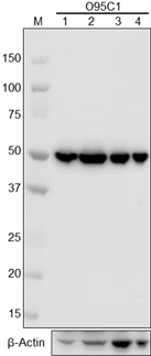
Western blot of purified anti-Histone H3 (C-terminus) antibody (clone 1B1-B2). Lane 1: Molecular weight marker; Lane 2: 20 µg of human brain lysate; Lane 3: 20 µg of mouse brain lysate; Lane 4: 20µg of rat brain lysate; Lane 5: 10µg of Drosophila head lysate. The blot was incubated with 0.1 µg/mL of the primary antibody overnight at 4°C, followed by incubation with HRP Goat anti-mouse IgG Antibody (Cat. No. 405306). Enhanced chemiluminescence was used as the detection system. -

Western blot of purified anti-Histone H3 (C-terminus) antibody (clone 1B1-B2). Lane 1: Molecular weight marker; Lane 2: 20 µg of Drosophila head lysate; Lane 3: 20 µg of Drosophila S2 (embryonic) cell lysate. The blot was incubated with 0.1 µg/mL of the primary antibody overnight at 4°C, followed by incubation with HRP Goat anti-mouse IgG Antibody (Cat. No. 405306). Enhanced chemiluminescence was used as the detection system.
| Cat # | Size | Price | Quantity Check Availability | Save | ||
|---|---|---|---|---|---|---|
| 819411 | 25 µg | 72€ | ||||
| 819412 | 100 µg | 216€ | ||||
Histone proteins are classified into core histones (H2A, H2B, H3, H4) and linker histones (H1, H5). Core histones form an octamer, which contains two H2A-H2B dimers and one H3-H4 tetramer. Core histones are predominantly globular except for the unstructured N-terminal tails. Posttranslational modifications, such as acetylation, methylation, phosphorylation, ubiquitination, SUMOylation and ADP-ribosylation occur in histone tails.
Histone modifications induce changes of chromatin structure and thereby affect the accessibility of transcription factors, nuclear proteins and enzymes to genomic DNA, resulting in gene activation or repression. It is known that histone modifications play critical roles in DNA repair, DNA replication, transcription regulation, alternative splicing and chromosome condensation and some diseases including autoimmune diseases and cancers.
Product Details
- Verified Reactivity
- Human, Mouse, Rat, Drosophila
- Antibody Type
- Monoclonal
- Host Species
- Mouse
- Immunogen
- This monoclonal antibody was raised against a peptide sequence derived from the C-terminal domain of human Histone H3 protein.
- Formulation
- 50 mM sodium citrate, 150mM sodium chloride, pH 5.8 with stabilizer
- Preparation
- The antibody was purified by affinity chromatography.
- Concentration
- 0.5 mg/mL
- Storage & Handling
- The antibody solution should be stored undiluted between 2°C and 8°C.
- Application
-
IHC-P - Quality tested
WB - Verified - Recommended Usage
-
Each lot of this antibody is quality control tested by immunohistochemistry. For immunohistochemistry, a concentration of 1.0 µg/mL is suggested. For Western blotting, the suggested use of this reagent is 0.01 - 0.5 µg/mL. It is recommended that the reagent be titrated for optimal performance for each application.
- Application Notes
-
Reactivity to Drosophila was only verified with the purified format.
- Additional Product Notes
-
For Western blotting, the suggested use of this reagent is 0.01 - 0.2 µg/mL in human, mouse, and rat and 0.1 - 0.5 µg/mL in Drosophila.
- Product Citations
-
- RRID
-
AB_2820127 (BioLegend Cat. No. 819411)
AB_2820128 (BioLegend Cat. No. 819412)
Antigen Details
- Structure
- Histone proteins H3 and H4 bind to form a H3-H4 dimer, two of these H3-H4 dimers combine to form a tetramer. This tetramer further combines with two H2a-H2b dimers to form the compact Histone octamer core.
- Distribution
-
Nucleus
- Function
- Histones play a central role in transcription regulation, DNA repair, DNA replication and chromosomal stability.
- Interaction
- Rsk-2, MSK1, GCN5 family of HATs, HDACs, PARP, SET7/9, and CARM1
- Biology Area
- Cell Biology, Chromatin Remodeling/Epigenetics, Neuroscience, Transcription Factors
- Antigen References
-
1. Chen HM, et al. 2015. Epigenetics and Dermatology 409.
- Gene ID
- 8350 View all products for this Gene ID
- UniProt
- View information about Histone on UniProt.org

Related FAQs
Other Formats
View All Histone Reagents Request Custom Conjugation| Description | Clone | Applications |
|---|---|---|
| Alexa Fluor® 594 anti-Histone H3 (C-terminus) | 1B1-B2 | ICC,IHC-F,IHC-P |
| HRP anti-Histone H3 (C-terminus) | 1B1-B2 | WB,IHC-P |
| Alexa Fluor® 647 anti-Histone H3 (C-terminus) | 1B1-B2 | IHC-P |
| Purified anti-Histone H3 (C-terminus) | 1B1-B2 | IHC-P,WB |
Customers Also Purchased
Compare Data Across All Formats
This data display is provided for general comparisons between formats.
Your actual data may vary due to variations in samples, target cells, instruments and their settings, staining conditions, and other factors.
If you need assistance with selecting the best format contact our expert technical support team.
-
Alexa Fluor® 594 anti-Histone H3 (C-terminus)

C57BL/6 mouse frozen testis section was fixed with 4% parafo... 
TE-71 cells were fixed with 1% paraformaldehyde (PFA), perme... 
Human paraffin-embedded breast cancer tissue slices were pre... 
Human paraffin-embedded colon cancer tissue slices were prep... 
Human paraffin-embedded colon tissue slices were prepared wi... -
HRP anti-Histone H3 (C-terminus)

Western blot of HRP anti-Histone H3 (C-terminus) antibody (c... 
IHC staining of HRP anti-Histone H3 (C-terminus) antibody (c... 
IHC staining of HRP anti-Histone H3 (C-terminus) antibody (c... 
IHC staining of HRP anti-Histone H3 (C-terminus) antibody (c... -
Alexa Fluor® 647 anti-Histone H3 (C-terminus)

IHC staining of Alexa Fluor® 647 anti-Histone H3 antibody (c... 
IHC staining of Alexa Fluor® 647 anti-Histone H3 antibody (c... 
IHC staining of Alexa Fluor® 647 anti-Histone H3 antibody (c... 
IHC staining of Alexa Fluor® 647 anti-Histone H3 antibody (c... -
Purified anti-Histone H3 (C-terminus)

IHC staining of purified anti-Histone H3 (C-Terminus) antibo... 
IHC staining of purified anti-Histone H3 (C-Terminus) antibo... 
IHC staining of purified anti-Histone H3 (C-Terminus) antibo... 
Western blot of purified anti-Histone H3 (C-terminus) antibo... 
Western blot of purified anti-Histone H3 (C-terminus) antibo...


Follow Us