 Login / Register


- Clone
- QBEnd/10 (See other available formats)
- Regulatory Status
- RUO
- Other Names
- Hematopoietic progenitor cell antigen CD34, CD34 antigen
- Previously
-
Signet Catalog# 326-26 L2 Predilute
Covance Catalog# SIG-3326
- Isotype
- Mouse IgG1, κ
- Ave. Rating
- Submit a Review
- Product Citations
- publications

-

Robust cytoplasmic and membrane staining of endothelial cells in frozen human placenta with antibody CD34. -

SeqIF™ (sequential immunofluorescence) staining on COMET™ of Purified anti-CD34 (clone QBEnd/10, yellow) on formalin-fixed paraffin-embedded human pancreatic carcinoma tissue at 1.25 µg/mL. Alexa Fluor™ Plus 647 Goat anti-Mouse IgG antibody (Lunaphore, Cat. No. DR647MS) was used as a secondary antibody. Nuclei were counterstained with DAPI (blue). Tissue underwent an all-in-one dewaxing and antigen retrieval preprocessing.
| Cat # | Size | Price | Quantity Check Availability | Save | ||
|---|---|---|---|---|---|---|
| 826401 | 50 µL | 168€ | ||||
CD34, also known as gp105-120, is a type I monomeric sialomucin-like glycophosphoprotein with an approximate molecular weight of 105-120 kD. Selectively expressed on the majority of hematopoietic stem/progenitor cells, bone marrow stromal cells, capillary endothelial cells, embryonic fibroblasts, and some nervous tissue, CD34 is a commonly used marker to identify human hematopoietic stem/progenitor cells. According to the differential sensitivity to enzymatic cleavage, four groups of epitopes of CD34 have been described. CD34 mediates cell adhesion and lymphocytes homing through binding to L-selectin and E-selectin ligands.
Product Details
Product Details
- Verified Reactivity
- Human
- Antibody Type
- Monoclonal
- Host Species
- Mouse
- Immunogen
- This antibody was generated using a vesicular suspension of a perfusate of human term placenta.
- Formulation
-
Phosphate-buffered solution with BSA + 0.09% NaN3.
Previous lots of this product may have been formulated with 0.1% or 0.05% NaN3 instead of 0.09% NaN3. For further information please contact BioLegend Technical Support or Customer Service. - Preparation
- The antibody was purified by affinity chromatography.
- Concentration
- 1.0 mg/ml
- Storage & Handling
- Store between 2°C and 8°C.
- Application
-
IHC - Quality tested
SB - Community verified - Recommended Usage
-
Each lot of this antibody is quality control tested by immunohistochemical staining.
The optimal working dilution should be determined for each specific assay condition.
• IHC: 1:1000-1:1500 (concentrated format) with Biotin based detection systems such as USA Ultra Streptavidin Detection (Cat. No. 929501).
Tissue Sections: Formalin-fixed, paraffin-embedded tissues, frozen sections
Pretreatment: For optimal staining, the sections should be pretreated with an antigen unmasking solution such as Retrieve-All 1 (Cat. No. 927901).
Incubation: 20 minutes at room temperature - Application Notes
-
This antibody is effective in immunohistochemistry (IHC).
The BioLegend QBEnd/10 monoclonal antibody detects the CD34 antigen. It stains the cytoplasm of endothelial cells of vascular tissue with some cross reactivity to basement membrane collagen. In tumors this monoclonal antibody is valuable in labelling the vascular endothelium of Kaposi's sarcoma, tumors involving the liver, spindle shaped tumors and any other highly vascular neoplastic or normal tissue.
**We recommend a quick spin prior to opening the vial.
Positive tissue (human): Skin, tonsil or other highly vascular tissue. - Additional Product Notes
-
For the use of this antibody in spatial biology applications, we have partnered with Lunaphore Technologies for demonstration of our antibodies on the COMET™. The COMET™ platform is an automated, end-to-end spatial biology solution developed for rapid and flexible multiplex tissue profiling. More information on the COMET™ and a complete list of our antibodies that have been demonstrated on the COMET™ can be found here.
-
Application References
(PubMed link indicates BioLegend citation) -
- Anthony P, et al. Endothelial markers in malignant vascular tumor of the liver: superiority of QBEnd/10 over von willebrand factor and uelex europus agglutinin 1. J Clin Path 44:2, 1991.
- Sankey EA, et al. QBEnd/10: a new immunostain for the diagnosis of Kaposi's Sarcoma. J Path 161:267-271, 1990.
- Fletcher CDM, et al. QBEnd/10: a useful but by no means a specific marker for the diagnosis of kaposi's sarcoma. J Path 162:274, 1990.
- Ramani P, et al. QBEnd/10, a new monoclonal antibody to endothelium: assessment of its diagnostic utility in paraffin sections. Histopathol 17:237-242, 1990.
- Product Citations
-
- RRID
-
AB_2564903 (BioLegend Cat. No. 826401)
Antigen Details
- Structure
- 105-120 kD single chain mucin-like glycoprotein
- Distribution
-
Hematopoietic stem/progenitor cells, bone marrow stromal cells, endothelial cells, embryonic fibroblasts
- Function
- Cell adhesion
- Ligand/Receptor
- L-selectin, E-selectin
- Cell Type
- Endothelial cells, Fibroblasts, Hematopoietic stem and progenitors
- Biology Area
- Cell Biology, Neuroinflammation, Neuroscience
- Antigen References
-
1. Krause DS, et al. 1996. Blood 87:1.
2. Puri KD, et al. 1995. J. Cell Biol. 131:261.
3. Zola H, et al. 2007. Leukocyte and Stromal Cell Molecules:The CD Markers. John Wiley & Sons Inc, Hoboken New Jersey. - Gene ID
- 947 View all products for this Gene ID
- UniProt
- View information about CD34 on UniProt.org

Related FAQs
- If an antibody clone has been previously successfully used in IBEX in one fluorescent format, will other antibody formats work as well?
-
It’s likely that other fluorophore conjugates to the same antibody clone will also be compatible with IBEX using the same sample fixation procedure. Ultimately a directly conjugated antibody’s utility in fluorescent imaging and IBEX may be specific to the sample and microscope being used in the experiment. Some antibody clone conjugates may perform better than others due to performance differences in non-specific binding, fluorophore brightness, and other biochemical properties unique to that conjugate.
- Will antibodies my lab is already using for fluorescent or chromogenic IHC work in IBEX?
-
Fundamentally, IBEX as a technique that works much in the same way as single antibody panels or single marker IF/IHC. If you’re already successfully using an antibody clone on a sample of interest, it is likely that clone will have utility in IBEX. It is expected some optimization and testing of different antibody fluorophore conjugates will be required to find a suitable format; however, legacy microscopy techniques like chromogenic IHC on fixed or frozen tissue is an excellent place to start looking for useful antibodies.
- Are other fluorophores compatible with IBEX?
-
Over 18 fluorescent formats have been screened for use in IBEX, however, it is likely that other fluorophores are able to be rapidly bleached in IBEX. If a fluorophore format is already suitable for your imaging platform it can be tested for compatibility in IBEX.
- The same antibody works in one tissue type but not another. What is happening?
-
Differences in tissue properties may impact both the ability of an antibody to bind its target specifically and impact the ability of a specific fluorophore conjugate to overcome the background fluorescent signal in a given tissue. Secondary stains, as well as testing multiple fluorescent conjugates of the same clone, may help to troubleshoot challenging targets or tissues. Using a reference control tissue may also give confidence in the specificity of your staining.
- How can I be sure the staining I’m seeing in my tissue is real?
-
In general, best practices for validating an antibody in traditional chromogenic or fluorescent IHC are applicable to IBEX. Please reference the Nature Methods review on antibody based multiplexed imaging for resources on validating antibodies for IBEX.
Other Formats
View All CD34 Reagents Request Custom Conjugation| Description | Clone | Applications |
|---|---|---|
| Purified anti-CD34 | QBEnd/10 | IHC,SB |
Customers Also Purchased
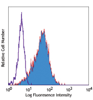
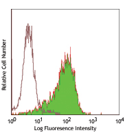
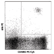
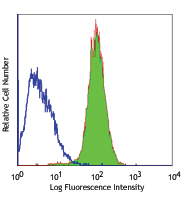
Compare Data Across All Formats
This data display is provided for general comparisons between formats.
Your actual data may vary due to variations in samples, target cells, instruments and their settings, staining conditions, and other factors.
If you need assistance with selecting the best format contact our expert technical support team.


Follow Us