 Login / Register


- Clone
- HNK-1 (See other available formats)
- Regulatory Status
- RUO
- Other Names
- HNK-1, NK-1, Leu-7
- Isotype
- Mouse IgM, κ
- Ave. Rating
- Submit a Review
- Product Citations
- publications

-

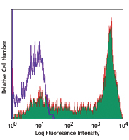
Human peripheral blood lymphocytes were stained with CD8 FITC and CD57 (clone HNK-1) PE (top) or mouse IgM, κ PE isotype control (bottom). -

| Cat # | Size | Price | Quantity Check Availability | Save | ||
|---|---|---|---|---|---|---|
| 359611 | 25 tests | 104€ | ||||
| 359612 | 100 tests | 249€ | ||||
CD57, also known as HNK-1, NK-1, and Leu-7 is a 100-115 kD oligosaccharide antigenic determinant expressed on a variety of proteins, lipids, and chondroitin sulfate proteoglycans. CD57 is expressed on a subset of peripheral blood lymphocytes, including NK cells and CD8+ T cells, and is also expressed on neural cells and striated muscle. CD57 is not expressed on red blood cells, granulocytes, monocytes, or platelets. While the function of CD57 is unknown, binding to L-selectin, P-selectin, and a fragment of laminin suggests that CD57 may be involved in cell-matrix interactions. CD57 is increased in some disease states associated with CD4/CD8 imbalances (AIDS, autoimmune disease, viral infections, and allograft transplants).
Product DetailsProduct Details
- Verified Reactivity
- Human
- Antibody Type
- Monoclonal
- Host Species
- Mouse
- Immunogen
- Membrane extract of human lymphoblastoid cell line HSB-2.
- Formulation
- Phosphate-buffered solution, pH 7.2, containing 0.09% sodium azide and BSA (origin USA)
- Preparation
- The antibody was purified by affinity chromatography and conjugated with PE under optimal conditions.
- Concentration
- Lot-specific (to obtain lot-specific concentration and expiration, please enter the lot number in our Certificate of Analysis online tool.)
- Storage & Handling
- The antibody solution should be stored undiluted between 2°C and 8°C, and protected from prolonged exposure to light. Do not freeze.
- Application
-
FC - Quality tested
SB - Community verified - Recommended Usage
-
Each lot of this antibody is quality control tested by immunofluorescent staining with flow cytometric analysis. For flow cytometric staining, the suggested use of this reagent is 5 µl per million cells in 100 µl staining volume or 5 µl per 100 µl of whole blood.
- Excitation Laser
-
Blue Laser (488 nm)
Green Laser (532 nm)/Yellow-Green Laser (561 nm)
- Application Notes
-
Additional reported applications for the relevant formats include: Western blotting1.
- Additional Product Notes
-
This product has been verified for IHC-P (Immunohistochemistry - formalin-fixed paraffin-embedded tissues) on the NanoString GeoMx® Digital Spatial Profiler. The GeoMx® enables researchers to perform spatial analysis of protein and RNA targets in FFPE and fresh frozen human and mouse samples. For more information about our spatial biology products and the GeoMx® platform, please visit our spatial biology page.
-
Application References
(PubMed link indicates BioLegend citation) -
- Yoshihara Y, et al. 1991. J. Cell Biol. 115:731. (WB)
- Abo T, et al. 1981. J. Immunol. 127:1024.
- Abo T, et al. 1982. J. Immunol. 129:1752.
- Abo T, et al. 1982. J. Immunol. 129:1758.
- Product Citations
-
- RRID
-
AB_2562758 (BioLegend Cat. No. 359611)
AB_2562759 (BioLegend Cat. No. 359612)
Antigen Details
- Structure
- Oligosaccharide antigenic determinant present on a variety of proteins, lipids, and chrondroitin sulfate proteoglycans, 100-115 kD. The antigen is conserved across species.
- Distribution
-
CD57 antigen is expressed on a subset of peripheral blood lymphocytes including a subset of NK cells and a subset of CD8+ T cells. Also expressed on neural cells and striated muscle.
- Function
- Unknown function, may be involved in cell-matrix interactions.
- Ligand/Receptor
- Binds to L-selectin and P-selectin in a calcium-dependent manner, also binds to second globular domain of E8 laminin fragment.
- Cell Type
- Lymphocytes, NK cells, T cells
- Biology Area
- Costimulatory Molecules, Immunology
- Molecular Family
- CD Molecules
- Antigen References
-
1. Schubert J, et al. 1989. In Leucocyte Typing IV (Knapp W, ed) Oxford University Press Oxford pp 711-714.
2. Palmer BE, et al. 2005. J. Immunol. 175:8415.
3. Schachner M, et al. 1995. Trends Neurosci. 18:183.
4. Wood KL, et al. 2005. Clin. Immunol. 117:294. - Gene ID
- 27087 View all products for this Gene ID
- UniProt
- View information about CD57 on UniProt.org
Related Pages & Pathways
Pathways


Related FAQs
- What type of PE do you use in your conjugates?
- We use R-PE in our conjugates.
- If an antibody clone has been previously successfully used in IBEX in one fluorescent format, will other antibody formats work as well?
-
It’s likely that other fluorophore conjugates to the same antibody clone will also be compatible with IBEX using the same sample fixation procedure. Ultimately a directly conjugated antibody’s utility in fluorescent imaging and IBEX may be specific to the sample and microscope being used in the experiment. Some antibody clone conjugates may perform better than others due to performance differences in non-specific binding, fluorophore brightness, and other biochemical properties unique to that conjugate.
- Will antibodies my lab is already using for fluorescent or chromogenic IHC work in IBEX?
-
Fundamentally, IBEX as a technique that works much in the same way as single antibody panels or single marker IF/IHC. If you’re already successfully using an antibody clone on a sample of interest, it is likely that clone will have utility in IBEX. It is expected some optimization and testing of different antibody fluorophore conjugates will be required to find a suitable format; however, legacy microscopy techniques like chromogenic IHC on fixed or frozen tissue is an excellent place to start looking for useful antibodies.
- Are other fluorophores compatible with IBEX?
-
Over 18 fluorescent formats have been screened for use in IBEX, however, it is likely that other fluorophores are able to be rapidly bleached in IBEX. If a fluorophore format is already suitable for your imaging platform it can be tested for compatibility in IBEX.
- The same antibody works in one tissue type but not another. What is happening?
-
Differences in tissue properties may impact both the ability of an antibody to bind its target specifically and impact the ability of a specific fluorophore conjugate to overcome the background fluorescent signal in a given tissue. Secondary stains, as well as testing multiple fluorescent conjugates of the same clone, may help to troubleshoot challenging targets or tissues. Using a reference control tissue may also give confidence in the specificity of your staining.
- How can I be sure the staining I’m seeing in my tissue is real?
-
In general, best practices for validating an antibody in traditional chromogenic or fluorescent IHC are applicable to IBEX. Please reference the Nature Methods review on antibody based multiplexed imaging for resources on validating antibodies for IBEX.
Other Formats
View All CD57 Reagents Request Custom Conjugation| Description | Clone | Applications |
|---|---|---|
| Purified anti-human CD57 | HNK-1 | FC,IHC-P,WB |
| FITC anti-human CD57 | HNK-1 | FC,SB |
| Pacific Blue™ anti-human CD57 | HNK-1 | FC |
| APC anti-human CD57 | HNK-1 | FC |
| PE anti-human CD57 | HNK-1 | FC,SB |
| Alexa Fluor® 647 anti-human CD57 | HNK-1 | FC,IHC-P,SB |
| Biotin anti-human CD57 | HNK-1 | FC |
| PE/Dazzle™ 594 anti-human CD57 | HNK-1 | FC |
| PerCP/Cyanine5.5 anti-human CD57 | HNK-1 | FC |
| PE/Cyanine7 anti-human CD57 | HNK-1 | FC |
| Alexa Fluor® 594 anti-human CD57 | HNK-1 | IHC-P,SB |
| Spark Blue™ 515 anti-human CD57 | HNK-1 | FC |
Customers Also Purchased
Compare Data Across All Formats
This data display is provided for general comparisons between formats.
Your actual data may vary due to variations in samples, target cells, instruments and their settings, staining conditions, and other factors.
If you need assistance with selecting the best format contact our expert technical support team.
-
Purified anti-human CD57

Human peripheral blood lymphocytes were stained with CD8 APC... 

Human paraffin-embedded cerebellum tissue slices were prepar... -
FITC anti-human CD57

Human peripheral blood lymphocytes were stained with CD8 APC... 
-
Pacific Blue™ anti-human CD57

Human peripheral blood lymphocytes were stained with CD8 APC... 
-
APC anti-human CD57

Human peripheral blood lymphocytes were stained with CD8 FIT... 
-
PE anti-human CD57

Human peripheral blood lymphocytes were stained with CD8 FIT... 
-
Alexa Fluor® 647 anti-human CD57


Human paraffin-embedded cerebellum tissue slices were prepar... -
Biotin anti-human CD57

Human peripheral blood lymphocytes were stained with CD8 FIT... 
-
PE/Dazzle™ 594 anti-human CD57

Human peripheral blood lymphocytes were stained with CD8 APC... -
PerCP/Cyanine5.5 anti-human CD57

Human peripheral blood lymphocytes were stained with CD8 APC... -
PE/Cyanine7 anti-human CD57

Human peripheral blood lymphocytes were stained with CD8 APC... -
Alexa Fluor® 594 anti-human CD57

Human paraffin-embedded cerebellum tissue slices were prepar... 
Human paraffin-embedded cerebellum tissue slices were prepar... -
Spark Blue™ 515 anti-human CD57

Human peripheral blood cells were surface stained with anti-...


Follow Us