 Login / Register


- Clone
- ME20.4 (See other available formats)
- Regulatory Status
- RUO
- Other Names
- p75NTR, TNFRSF16, p75(NTR), Gp80-LNGFR, NGFR, Nerve Growth Factor Receptor
- Isotype
- Mouse IgG1, κ
- Ave. Rating
- Submit a Review
- Product Citations
- publications

-

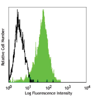
Human neuroblastoma cell line SK-N-MC stained with ME20.4 PE
| Cat # | Size | Price | Quantity Check Availability | Save | ||
|---|---|---|---|---|---|---|
| 345105 | 25 tests | 123€ | ||||
| 345106 | 100 tests | 244€ | ||||
CD271, also known as p75NTR, TNFRSF16, p75(NTR), Gp80-LNGFR, and NGFR, is a type I transmembrane protein with a MW of 75 kD. It is expressed by many cell types including neurons, Schwann cells, mesenchymal stem/stromal cells, follicular dendritic cells and melanocytes. The extracellular portion contains four TNFR-Cys repeats that form the binding domain for its ligands (NGF, BDNF, NTF3, and NTF4). The intracellular portion of CD271 contains a death domain, which interacts with TRAF2, TRAF4, TRAF6, PTPN13 and RANBP9, to promote cell apoptosis, and to regulate cell differentiation and neurogenesis.
Product DetailsProduct Details
- Verified Reactivity
- Human
- Reported Reactivity
- African Green, Baboon, Cynomolgus, Rhesus
- Antibody Type
- Monoclonal
- Host Species
- Mouse
- Immunogen
- WM245 melanoma cells
- Formulation
- Phosphate-buffered solution, pH 7.2, containing 0.09% sodium azide and BSA (origin USA)
- Preparation
- The antibody was purified by affinity chromatography, and conjugated with PE under optimal conditions.
- Concentration
- Lot-specific (to obtain lot-specific concentration and expiration, please enter the lot number in our Certificate of Analysis online tool.)
- Storage & Handling
- The antibody solution should be stored undiluted between 2°C and 8°C, and protected from prolonged exposure to light. Do not freeze.
- Application
-
FC - Quality tested
- Recommended Usage
-
Each lot of this antibody is quality control tested by immunofluorescent staining with flow cytometric analysis. For flow cytometric staining, the suggested use of this reagent is 5 µl per million cells in 100 µl staining volume or 5 µl per 100 µl of whole blood.
- Excitation Laser
-
Blue Laser (488 nm)
Green Laser (532 nm)/Yellow-Green Laser (561 nm)
- Application Notes
-
Additional reported applications for the relevant formats include: immunoprecipitation1, Western blotting4, immunohistochemical staining of frozen or paraffin embedded tissue sections1,2, and blocking of NGF binding1.
-
Application References
(PubMed link indicates BioLegend citation) -
- Ross A, et al. 1984. P. Natl. Acad. Sci. USA 81:6681. (FC, IP, Block, IHC)
- Cattoretti G, et al. 1993. Blood 81:1726. (IHC)
- Kamke MR, et al. 2005. Hear Res. 206:89.
- Baker D, et al. 1989. Cancer Res. 49:4142-4146. (FC, WB)
- Product Citations
-
- RRID
-
AB_2282827 (BioLegend Cat. No. 345105)
AB_2152647 (BioLegend Cat. No. 345106)
Antigen Details
- Structure
- Type I transmembranal protein with a MW of 75 kD. The extracellular portion contains four TNFR-Cys repeats that form the NGF binding domain; the intracellular portion contains a death domain.
- Distribution
-
Expressed by many cell types including neurons, Schwann cells, mesenchymal stem/stromal cells, follicular dendritic cells, melanocytes.
- Function
- Apoptosis, differentiation, neurogenesis.
- Interaction
- TRAF2, TRAF4, TRAF6, PTPN13, RANBP9.
- Ligand/Receptor
- Nerve growth factor (NGF), Brain-derived neurotrophic factor (BDNF), Neurotrophin 3. (NTF3) , Neurotrophin 4 (NTF4)
- Bioactivity
- Apoptosis, differentiation, neurogenesis.
- Cell Type
- Dendritic cells, Mesenchymal cells, Mesenchymal Stem Cells, Neurons
- Biology Area
- Apoptosis/Tumor Suppressors/Cell Death, Cell Biology, Immunology, Neuroscience, Stem Cells, Synaptic Biology
- Molecular Family
- CD Molecules, Cytokine/Chemokine Receptors, Postsynaptic proteins
- Antigen References
-
1. Friedman MJ, et al. 2009. J. Biol. Chem. 284:27944.
2. Rock JR, et al. 2009. Proc Natl Acad Sci USA. 106:12771.
3. Kidd SK, et al. 2008. Brain Res.1195:113.
4. Peters EM, et al. 2007. Am J Pathol. 171:1872.
5. Jansen P, et al. 2007. Nat Neurosci. 10:1449. - Gene ID
- 4804 View all products for this Gene ID
- UniProt
- View information about CD271 on UniProt.org
Related Pages & Pathways
Pathways


Related FAQs
- What type of PE do you use in your conjugates?
- We use R-PE in our conjugates.
Other Formats
View All CD271 Reagents Request Custom Conjugation| Description | Clone | Applications |
|---|---|---|
| Purified anti-human CD271 (NGFR) | ME20.4 | FC,IP,WB,IHC-P,IHC-F |
| FITC anti-human CD271 (NGFR) | ME20.4 | FC |
| PE anti-human CD271 (NGFR) | ME20.4 | FC |
| APC anti-human CD271 (NGFR) | ME20.4 | FC |
| PE/Cyanine7 anti-human CD271 (NGFR) | ME20.4 | FC |
| PerCP/Cyanine5.5 anti-human CD271 (NGFR) | ME20.4 | FC |
| Alexa Fluor® 647 anti-human CD271 (NGFR) | ME20.4 | FC |
| Alexa Fluor® 700 anti-human CD271 (NGFR) | ME20.4 | FC |
| PE/Dazzle™ 594 anti-human CD271 (NGFR) | ME20.4 | FC |
| APC/Fire™ 750 anti-human CD271 (NGFR) | ME20.4 | FC |
| Biotin anti-human CD271 (NGFR) | ME20.4 | FC |
| TotalSeq™-A0816 anti-human CD271 (NGFR) | ME20.4 | PG |
| APC/Cyanine7 anti-human CD271 (NGFR) | ME20.4 | FC |
| TotalSeq™-C0816 anti-human CD271 (NGFR) | ME20.4 | PG |
| TotalSeq™-B0816 anti-human CD271 (NGFR) | ME20.4 | PG |
| Pacific Blue™ anti-human CD271 (NGFR) Antibody | ME20.4 | FC |
| TotalSeq™-D0816 anti-human CD271 (NGFR) | ME20.4 | PG |
Customers Also Purchased
Compare Data Across All Formats
This data display is provided for general comparisons between formats.
Your actual data may vary due to variations in samples, target cells, instruments and their settings, staining conditions, and other factors.
If you need assistance with selecting the best format contact our expert technical support team.
-
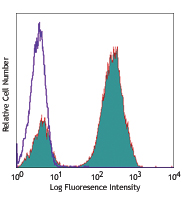
Purified anti-human CD271 (NGFR)

Human neuroblastoma cell line SK-N-MC stained with ME20.4 FI... -
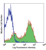
FITC anti-human CD271 (NGFR)
Human neuroblastoma cell line SK-N-MC stained with ME20.4 FI... -
PE anti-human CD271 (NGFR)

Human neuroblastoma cell line SK-N-MC stained with ME20.4 PE -
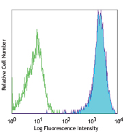
APC anti-human CD271 (NGFR)

Human neuroblastoma cell line, SK-N-MC, stained with ME20.4 ... -
PE/Cyanine7 anti-human CD271 (NGFR)

Human neuroblastoma cell line, SK-N-MC, was stained with CD2... -
PerCP/Cyanine5.5 anti-human CD271 (NGFR)

Human neuroblastoma cell line, SK-N-MC, was stained with CD2... -
Alexa Fluor® 647 anti-human CD271 (NGFR)

Human neuroblastoma cell line, SK-N-MC, was stained with CD2... -
Alexa Fluor® 700 anti-human CD271 (NGFR)

Human neuroblastoma cell line, SK-N-MC, was stained with CD2... -
PE/Dazzle™ 594 anti-human CD271 (NGFR)

Human neuroblastoma cell line, SK-N-MC, was stained with CD2... -
APC/Fire™ 750 anti-human CD271 (NGFR)

Human neuroblastoma cell line, SK-N-MC, was stained with CD2... -
Biotin anti-human CD271 (NGFR)

Human neuroblastoma cell line SK-N-MC was stained with bioti... -
TotalSeq™-A0816 anti-human CD271 (NGFR)
-
APC/Cyanine7 anti-human CD271 (NGFR)

Human neuroblastoma cell line, SK-N-MC, was stained with CD2... -
TotalSeq™-C0816 anti-human CD271 (NGFR)
-
TotalSeq™-B0816 anti-human CD271 (NGFR)
-
Pacific Blue™ anti-human CD271 (NGFR) Antibody

Human neuroblastoma cell line, SK-N-MC, was stained with ant... -
TotalSeq™-D0816 anti-human CD271 (NGFR)


Follow Us