 Login / Register


- Clone
- 2A9-1 (See other available formats)
- Regulatory Status
- RUO
- Other Names
- V28, GPR13, Chemokine C-X3-C receptor 1
- Isotype
- Rat IgG2b, κ
- Ave. Rating
- Submit a Review
- Product Citations
- publications

-

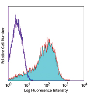
Human peripheral blood lymphocytes were stained with CD3 PE and CX3CR1 (clone 2A9-1) Brilliant Violet 650™ (top) or rat IgG2b, κ isotype control (bottom). -

| Cat # | Size | Price | Quantity Check Availability | Save | ||
|---|---|---|---|---|---|---|
| 341625 | 25 tests | 168€ | ||||
| 341626 | 100 tests | 326€ | ||||
CX3CR1 is a G-protein-coupled seven-transmembrane chemokine receptor, also called GPR13 or V28. It is expressed on NK cells, T cell subset, monocytes/macrophages, dendritic cells, and some malignant epithelial cells. CX3CL1 (known also as fractalkine and neurotactin) is the ligand of CX3CR1. CX3CL1 is a unique transmembrane molecule with a CX3C-motif chemokine domain and a mucin-like stalk. CX3CL1 is expressed by activated-endothelial cells, neurons, and astrocytes. The interaction of CX3CR1 and its ligand mediatesfirm cell adhesion and migration.
Product DetailsProduct Details
- Verified Reactivity
- Human
- Reported Reactivity
- African Green, Cynomolgus, Rhesus
- Antibody Type
- Monoclonal
- Host Species
- Rat
- Immunogen
- CX3CR1-EGFP fusion protein
- Formulation
- Phosphate-buffered solution, pH 7.2, containing 0.09% sodium azide and BSA (origin USA).
- Preparation
- The antibody was purified by affinity chromatography and conjugated with Brilliant Violet 650™ under optimal conditions.
- Concentration
- Lot-specific (to obtain lot-specific concentration and expiration, please enter the lot number in our Certificate of Analysis online tool.)
- Storage & Handling
- The antibody solution should be stored undiluted between 2°C and 8°C, and protected from prolonged exposure to light. Do not freeze.
- Application
-
FC - Quality tested
- Recommended Usage
-
Each lot of this antibody is quality control tested by immunofluorescent staining with flow cytometric analysis. For flow cytometric staining, the suggested use of this reagent is 5 µl per million cells in 100 µl staining volume or 5 µl per 100 µl of whole blood.
Brilliant Violet 650™ excites at 405 nm and emits at 645 nm. The bandpass filter 660/20 nm is recommended for detection, although filter optimization may be required depending on other fluorophores used. Be sure to verify that your cytometer configuration and software setup are appropriate for detecting this channel. Refer to your instrument manual or manufacturer for support. Brilliant Violet 650™ is a trademark of Sirigen Group Ltd.
Learn more about Brilliant Violet™.
This product is subject to proprietary rights of Sirigen Inc. and is made and sold under license from Sirigen Inc. The purchase of this product conveys to the buyer a non-transferable right to use the purchased product for research purposes only. This product may not be resold or incorporated in any manner into another product for resale. Any use for therapeutics or diagnostics is strictly prohibited. This product is covered by U.S. Patent(s), pending patent applications and foreign equivalents. - Excitation Laser
-
Violet Laser (405 nm)
-
Application References
(PubMed link indicates BioLegend citation) -
- Nishimura M, et al. 2002. J. Immunol. 168:6173.
- Nanki T, et al. 2002. Arthritis Rheum. 46:2878.
- Kobayashi T, et al. 2007. Inflamm. Bowel Dis. 13:837.
- Beziat V, et al. 2011. J. Immunol. 186:6753. PubMed.
- Product Citations
-
- RRID
-
AB_2716244 (BioLegend Cat. No. 341625)
AB_2716245 (BioLegend Cat. No. 341626)
Antigen Details
- Structure
- G-protein-coupled seven transmembrane receptor
- Distribution
-
NK cells, T cell subset, monocytes/macrophages, dendritic cells
- Function
- cell adhesion and migration
- Ligand/Receptor
- CX3CL1 (Fractalkine, FKN, neurotactin)
- Cell Type
- Dendritic cells, Macrophages, Monocytes, NK cells, T cells
- Biology Area
- Cell Biology, Immunology, Neuroinflammation, Neuroscience, Neuroscience Cell Markers
- Molecular Family
- Cytokine/Chemokine Receptors, GPCR
- Antigen References
-
1. Imai T, et al. 1997. Cell. 91:521.
2. Fong AM, et al. 1998. J. Exp. Med. 188:1413.
3. Auffray C, et al. 2009. J. Exp. Med. 206:595. - Gene ID
- 1524 View all products for this Gene ID
- UniProt
- View information about CX3CR1 on UniProt.org
Related Pages & Pathways
Pathways


Related FAQs
- Does staining at room temperature or even at 37°C help for checking chemokine receptors expression?
-
Due to continuous recycling of many chemokine receptors, it may be worthwhile to consider staining at room temperature or at 37°C if the staining at lower temperature (which can potentially reduce receptor turnover) is not optimal.
Other Formats
View All CX3CR1 Reagents Request Custom Conjugation| Description | Clone | Applications |
|---|---|---|
| PE/Cyanine7 anti-human CX3CR1 | 2A9-1 | FC |
| Purified anti-human CX3CR1 | 2A9-1 | FC |
| PE anti-human CX3CR1 | 2A9-1 | FC |
| FITC anti-human CX3CR1 | 2A9-1 | FC |
| Alexa Fluor® 647 anti-human CX3CR1 | 2A9-1 | FC |
| APC anti-human CX3CR1 | 2A9-1 | FC |
| PerCP/Cyanine5.5 anti-human CX3CR1 | 2A9-1 | FC |
| APC/Cyanine7 anti-human CX3CR1 | 2A9-1 | FC |
| Biotin anti-human CX3CR1 | 2A9-1 | FC |
| Brilliant Violet 510™ anti-human CX3CR1 | 2A9-1 | FC |
| Brilliant Violet 421™ anti-human CX3CR1 | 2A9-1 | FC |
| PE/Dazzle™ 594 anti-human CX3CR1 | 2A9-1 | FC |
| Brilliant Violet 650™ anti-human CX3CR1 | 2A9-1 | FC |
| Brilliant Violet 785™ anti-human CX3CR1 | 2A9-1 | FC |
| Brilliant Violet 711™ anti-human CX3CR1 | 2A9-1 | FC |
| APC/Fire™ 750 anti-human CX3CR1 | 2A9-1 | FC |
| Brilliant Violet 570™ anti-human CX3CR1 | 2A9-1 | FC |
| Spark Red™ 718 anti-human CX3CR1 (Flexi-Fluor™) | 2A9-1 | FC |
Customers Also Purchased

Compare Data Across All Formats
This data display is provided for general comparisons between formats.
Your actual data may vary due to variations in samples, target cells, instruments and their settings, staining conditions, and other factors.
If you need assistance with selecting the best format contact our expert technical support team.
-
PE/Cyanine7 anti-human CX3CR1

Human peripheral blood lymphocytes were stained with CD3 FIT... 
-
Purified anti-human CX3CR1

Human peripheral blood lymphocytes stained with CD3 APC (UCH... -
PE anti-human CX3CR1
Human peripheral blood lymphocytes stained with CD3 APC (UCH... -
FITC anti-human CX3CR1

Human peripheral blood lymphocytes stained with 2A9-1 FITC a... -
Alexa Fluor® 647 anti-human CX3CR1

Human peripheral blood lymphocytes stained with 2A9-1 Alexa ... -
APC anti-human CX3CR1

Human peripheral blood lymphocytes stained with CD3 FITC and... 
-
PerCP/Cyanine5.5 anti-human CX3CR1

Human peripheral blood lymphocytes were stained with CD3 FIT... -
APC/Cyanine7 anti-human CX3CR1

Human peripheral blood lymphocytes were stained with CD3 FIT... -
Biotin anti-human CX3CR1

Human peripheral blood lymphocytes stained with CD3 FITC and... 
-
Brilliant Violet 510™ anti-human CX3CR1

Human peripheral blood lymphocytes were stained with CD3 APC... 
-
Brilliant Violet 421™ anti-human CX3CR1

Human peripheral blood lymphocytes were stained with CD3 APC... 
-
PE/Dazzle™ 594 anti-human CX3CR1

Human peripheral blood lymphocytes were stained with CD3 APC... 
-
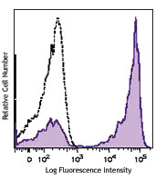
Brilliant Violet 650™ anti-human CX3CR1

Human peripheral blood lymphocytes were stained with CD3 PE ... 
-
Brilliant Violet 785™ anti-human CX3CR1

Human peripheral blood lymphocytes were stained with CD3 PE ... -
Brilliant Violet 711™ anti-human CX3CR1

Human peripheral blood lymphocytes were stained with CD3 PE ... -
APC/Fire™ 750 anti-human CX3CR1

Human peripheral blood lymphocytes were stained with CD3 PE ... -
Brilliant Violet 570™ anti-human CX3CR1

Human peripheral blood lymphocytes were stained with anti-hu... -
Spark Red™ 718 anti-human CX3CR1 (Flexi-Fluor™)


Follow Us