 Login / Register


- Clone
- AFS98 (See other available formats)
- Regulatory Status
- RUO
- Other Names
- CSF-1R, M-CSFR, c-fms
- Isotype
- Rat IgG2a, κ
- Ave. Rating
- Submit a Review
- Product Citations
- publications

-

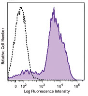
Thioglycolate-elicited C57BL/6 mouse peritoneal macrophages were stained with F4/80 APC and CD115 (clone AFS98) Brilliant Violet 605™ (top) or rat IgG2a, κ Brilliant Violet 605™ isotype control (bottom). -

| Cat # | Size | Price | Quantity Check Availability | Save | ||
|---|---|---|---|---|---|---|
| 135517 | 50 µg | 253€ | ||||
CSF-1R, also known as CD115 and M-CSFR, is a single-pass type I membrane protein and member of the platelet-derived growth factor receptor family. This c-fms (Fms proto-oncogene) gene product's natural ligands include M-CSF and IL-34. Structural studies of CD115 have described an Ig-like extracellular domain, a transmembrane domain, an intracellular juxtamembrane domain, a split tyrosine kinase domain, and a C-terminal tail receptor. Receptor activation induces homodimerization in addition to phosphorylation and ubiquitination of intracellular residues. CD115 directly influences tissue macrophage and osteoclast differentiation and proliferation. It is expressed on monocytes/macrophages, peritoneal exudate cells, plasmacytoid and conventional dendritic cells, and osteoclasts.
Product DetailsProduct Details
- Verified Reactivity
- Mouse
- Antibody Type
- Monoclonal
- Host Species
- Rat
- Formulation
- Phosphate-buffered solution, pH 7.2, containing 0.09% sodium azide and BSA (origin USA).
- Preparation
- The antibody was purified by affinity chromatography and conjugated with Brilliant Violet 605™ under optimal conditions.
- Concentration
- 0.2 mg/ml
- Storage & Handling
- The antibody solution should be stored undiluted between 2°C and 8°C, and protected from prolonged exposure to light. Do not freeze.
- Application
-
FC - Quality tested
- Recommended Usage
-
Each lot of this antibody is quality control tested by immunofluorescent staining with flow cytometric analysis. For flow cytometric staining, the suggested use of this reagent is ≤0.5 µg per million cells in 100 µl volume. It is recommended that the reagent be titrated for optimal performance for each application.
Brilliant Violet 605™ excites at 405 nm and emits at 603 nm. The bandpass filter 610/20 nm is recommended for detection, although filter optimization may be required depending on other fluorophores used. Be sure to verify that your cytometer configuration and software setup are appropriate for detecting this channel. Refer to your instrument manual or manufacturer for support. Brilliant Violet 605™ is a trademark of Sirigen Group Ltd.
Learn more about Brilliant Violet™.
This product is subject to proprietary rights of Sirigen Inc. and is made and sold under license from Sirigen Inc. The purchase of this product conveys to the buyer a non-transferable right to use the purchased product for research purposes only. This product may not be resold or incorporated in any manner into another product for resale. Any use for therapeutics or diagnostics is strictly prohibited. This product is covered by U.S. Patent(s), pending patent applications and foreign equivalents. - Excitation Laser
-
Violet Laser (405 nm)
- Application Notes
-
Additional reported applications (for the relevant formats) include: blocking of ligand binding1. The LEAF™ purified antibody (Endotoxin <0.1 EU/µg, Azide-Free, 0.2 µm filtered) is recommended for functional assays.
It has been reported that CD115 can be rapidly internalized, especially when samples are exposed to room temperature. Approximate 33% decrease in CD115 expression has been observed between 0 and 4 hours after sample collection, while overnight incubation of the cells results in complete CD115 downregulation. Pre-treatment with EDTA and low temperatures (2 to 8°C) helps in maintaining surface expression of CD1154. -
Application References
(PubMed link indicates BioLegend citation) - Product Citations
-
- RRID
-
AB_2562760 (BioLegend Cat. No. 135517)
Antigen Details
- Structure
- Ig superfamily, 145 kD
- Distribution
-
Monocytes/macrophages, peritoneal exudate cells, plasmacytoid and conventional dendritic cells, osteoclasts
- Function
- Growth factor receptor, tyrosine kinase
- Ligand/Receptor
- Macrophage colony stimulating factor (M-CSF), IL-34
- Cell Type
- Dendritic cells, Macrophages, Monocytes, Osteoclasts
- Biology Area
- Immunology
- Molecular Family
- CD Molecules, Cytokine/Chemokine Receptors
- Antigen References
-
- Sudo T, et al. 1995 Oncogene 11:2469.
- Murayama T, et al. 1999 Circulation 99:1740.
- Goswami S, et al. 2005 Cancer Res. 65:5278.
- Yu W, et al. 2008 J. Leuko. Biol. 84:852.
- Gene ID
- 12978 View all products for this Gene ID
- UniProt
- View information about CD115 on UniProt.org
Related Pages & Pathways
Pathways


Related FAQs
- Why do I have a weak CD115 staining?
-
It has been reported that CD115 can be rapidly internalized, especially when samples are exposed to room temperature. Approximate 33% decrease in CD115 expression has been observed between 0 and 4 hours after sample collection, while overnight incubation of the cells results in complete CD115 downregulation. Pre-treatment with EDTA and low temperatures (2 - 8°C) helps in maintaining surface expression of CD115. In addition, brief fixation of the cells with Fixation Buffer (Cat. No. 420801) for 10 minutes will block CD115 internalization.
Other Formats
View All CD115 Reagents Request Custom ConjugationCustomers Also Purchased
Compare Data Across All Formats
This data display is provided for general comparisons between formats.
Your actual data may vary due to variations in samples, target cells, instruments and their settings, staining conditions, and other factors.
If you need assistance with selecting the best format contact our expert technical support team.
-
Purified anti-mouse CD115 (CSF-1R)

Thioglycolate-elicited BALB/c mouse peritoneal macrophages s... 
Thioglycolate-elicited BALB/c mouse peritoneal macrophages s... -
PE anti-mouse CD115 (CSF-1R)
Thioglycolate-elicited BALB/c mouse peritoneal macrophages s...
Thioglycolate-elicited BALB/c mouse peritoneal macrophages s... -
Biotin anti-mouse CD115 (CSF-1R)

Thioglycolate-elicited BALB/c mouse peritoneal macrophages s... 
-
APC anti-mouse CD115 (CSF-1R)

Thioglycolate-elicted BALB/c mouse peritoneal macrophages st... 
-
Alexa Fluor® 488 anti-mouse CD115 (CSF-1R)

Thioglycolate-elicited C57BL/6 mouse peritoneal macrophages ... -
Brilliant Violet 421™ anti-mouse CD115 (CSF-1R)

Thioglycolate-elicited C57BL/6 mouse peritoneal macrophages ... 
-
Brilliant Violet 605™ anti-mouse CD115 (CSF-1R)

Thioglycolate-elicited C57BL/6 mouse peritoneal macrophages ... 
-
Brilliant Violet 711™ anti-mouse CD115 (CSF-1R)

Thioglycolate-elicited C57BL/6 mouse peritoneal macrophages ... 
-
Alexa Fluor® 594 anti-mouse CD115 (CSF-1R)

C57BL/6 mouse bone marrow derived macrophages were fixed wit... -
Purified anti-mouse CD115 (CSF-1R) (Maxpar® Ready)

Mouse J774A.1 macrophage cells (top) and mouse EL4 T cells (... -
PE/Cyanine7 anti-mouse CD115 (CSF-1R)

Thioglycolate-elicited BALB/c peritoneal macrophages were st... -
PerCP/Cyanine5.5 anti-mouse CD115 (CSF-1R)

Thioglycolate-elicited BALB/c peritoneal macrophages were st... -
PE/Dazzle™ 594 anti-mouse CD115 (CSF-1R)

Thioglycolate-elicited BALB/c mouse peritoneal macrophages w... 
-
Alexa Fluor® 647 anti-mouse CD115 (CSF-1R)

Thioglycolate-elicited BALB/c mouse peritoneal macrophages w... 
-
APC/Cyanine7 anti-mouse CD115 (CSF-1R)

Thioglycolate-elicited BALB/c mouse peritoneal macrophages w... -
TotalSeq™-A0105 anti-mouse CD115 (CSF-1R)
-
APC/Fire™ 750 anti-mouse CD115 (CSF-1R)

Thioglycolate-elicited C57BL/6 mouse peritoneal macrophages ... -
Ultra-LEAF™ Purified anti-mouse CD115 (CSF-1R)
-
TotalSeq™-B0105 anti-mouse CD115 (CSF-1R)
-
TotalSeq™-C0105 anti-mouse CD115 (CSF-1R)
-
Brilliant Violet 785™ anti-mouse CD115 (CSF-1R)

Thioglycolate-elicited BALB/c peritoneal macrophages were st...


Follow Us