 Login / Register


- Clone
- G10F5 (See other available formats)
- Regulatory Status
- RUO
- Workshop
- VI MA81
- Other Names
- CD67, CGM6, NCA-95, CEACAM8
- Isotype
- Mouse IgM, κ
- Ave. Rating
- Submit a Review
- Product Citations
- publications

-

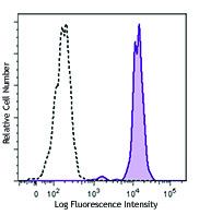
Human peripheral blood granulocytes were stained with CD66b (clone G10F5) APC/Cyanine7 (filled histogram) open histogram depicts unstained cells.
| Cat # | Size | Price | Quantity Check Availability | Save | ||
|---|---|---|---|---|---|---|
| 305125 | 25 tests | 123€ | ||||
| 305126 | 100 tests | 300€ | ||||
CD66b is a 95-100 kD glycosylphosphatidylinositol (GPI)-linked protein also known as CD67, CGM6, and NCA-95. CD66b is a member of the immunoglobulin superfamily, carcinoembryonic antigen (CEA)-like subfamily. CD66b, expressed on granulocytes, has been reported to induce activation in neutrophils and to be involved in heterophilic adhesion with CD66c.
Product DetailsProduct Details
- Verified Reactivity
- Human
- Reported Reactivity
- Chimpanzee
- Antibody Type
- Monoclonal
- Host Species
- Mouse
- Formulation
- Phosphate-buffered solution, pH 7.2, containing 0.09% sodium azide and BSA (origin USA)
- Preparation
- The antibody was purified by affinity chromatography and conjugated with APC/Cyanine7 under optimal conditions.
- Concentration
- Lot-specific (to obtain lot-specific concentration and expiration, please enter the lot number in our Certificate of Analysis online tool.)
- Storage & Handling
- The antibody solution should be stored undiluted between 2°C and 8°C, and protected from prolonged exposure to light. Do not freeze.
- Application
-
FC - Quality tested
- Recommended Usage
-
Each lot of this antibody is quality control tested by immunofluorescent staining with flow cytometric analysis. For flow cytometric staining, the suggested use of this reagent is 5 µl per million cells in 100 µl staining volume or 5 µl per 100 µl of whole blood.
- Excitation Laser
-
Red Laser (633 nm)
- Application Notes
-
Additional reported applications (for the relevant formats) include: immunohistochemical staining of acetone-fixed frozen, formalin-fixed paraffin-embedded tissue sections, and spatial biology (IBEX)5,6.
- Additional Product Notes
- BioLegend is in the process of converting the name APC/Cy7 to APC/Cyanine7. The dye molecule remains the same, so you should expect the same quality and performance from our APC/Cyanine7 products. Please contact Technical Service if you have any questions.
-
Application References
(PubMed link indicates BioLegend citation) -
- Schlossman S, et al. Eds. 1995. Leucocyte Typing V. Oxford University Press. New York.
- Kishimoto T, et al. Eds. 1997. Leucocyte Typing VI. Garland Publishing Inc. London.
- Norling LV, et al. 2012. Arterioscler Thromb Vasc Biol. 32:1970. PubMed
- Meinke P, et al. 2015. Neuroimmunol Discord. 25:127. PubMed
- Radtke AJ, et al. 2020. Proc Natl Acad Sci USA. 117:33455-33465. (SB) PubMed
- Radtke AJ, et al. 2022. Nat Protoc. 17:378-401. (SB) PubMed
- Product Citations
-
- RRID
-
AB_2750183 (BioLegend Cat. No. 305125)
AB_2750184 (BioLegend Cat. No. 305126)
Antigen Details
- Structure
- Ig superfamily, CEA antigen group, GPI-linked glycoprotein, 95-100 kD
- Distribution
-
Granulocytes
- Function
- Cell adhesion, neutrophil activation
- Ligand/Receptor
- CD66c
- Cell Type
- Granulocytes, Neutrophils
- Biology Area
- Immunology
- Molecular Family
- Adhesion Molecules, CD Molecules
- Antigen References
-
1. Kuijpers T, et al. 1993. J. Immunol. 151:4934.
2. Kuroki M, et al. 1992. J. Leuk. Biol. 52:551. - Gene ID
- 1088 View all products for this Gene ID
- UniProt
- View information about CD66b on UniProt.org
Related Pages & Pathways
Pathways


Related FAQs
Other Formats
View All CD66b Reagents Request Custom Conjugation| Description | Clone | Applications |
|---|---|---|
| FITC anti-human CD66b | G10F5 | FC |
| Purified anti-human CD66b | G10F5 | FC,IHC-P |
| Pacific Blue™ anti-human CD66b | G10F5 | FC |
| PE anti-human CD66b | G10F5 | FC |
| PerCP/Cyanine5.5 anti-human CD66b | G10F5 | FC |
| Alexa Fluor® 647 anti-human CD66b | G10F5 | FC,IHC-P,SB |
| Alexa Fluor® 700 anti-human CD66b | G10F5 | FC |
| PE/Cyanine7 anti-human CD66b | G10F5 | FC |
| APC anti-human CD66b | G10F5 | FC |
| Biotin anti-human CD66b | G10F5 | FC |
| PE/Dazzle™ 594 anti-human CD66b | G10F5 | FC |
| Alexa Fluor® 594 anti-human CD66b | G10F5 | IHC-P |
| APC/Cyanine7 anti-human CD66b | G10F5 | FC |
| FITC anti-human CD66b | G10F5 | FC |
| GMP FITC anti-human CD66b | G10F5 | FC |
Customers Also Purchased
Compare Data Across All Formats
This data display is provided for general comparisons between formats.
Your actual data may vary due to variations in samples, target cells, instruments and their settings, staining conditions, and other factors.
If you need assistance with selecting the best format contact our expert technical support team.
-
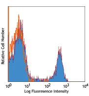
FITC anti-human CD66b

Human peripheral whole blood granulocytes stained with G10F5... -
Purified anti-human CD66b

Human peripheral blood granulocytes stained with purified G1... 
Human paraffin-embedded spleen tissue slices were prepared w... -
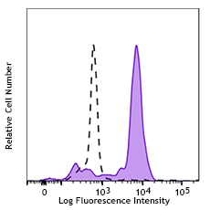
Pacific Blue™ anti-human CD66b

Human peripheral blood granulocytes were stained with CD66b ... -
PE anti-human CD66b

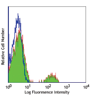
Human peripheral blood granulocytes stained with G10F5 PE -
PerCP/Cyanine5.5 anti-human CD66b

Human peripheral blood granulocytes were stained with CD66b ... -
Alexa Fluor® 647 anti-human CD66b

Human peripheral blood granulocytes were stained with CD66b ... 
Human paraffin-embedded spleen tissue was stained with Alexa... 
Confocal image of human lymph node sample acquired using the... -
Alexa Fluor® 700 anti-human CD66b

Human peripheral blood granulocytes were stained with CD66b ... -
PE/Cyanine7 anti-human CD66b

Human peripheral blood granulocytes were stained with CD66b ... -
APC anti-human CD66b

Human peripheral blood granulocytes were stained with CD66b ... -
Biotin anti-human CD66b

Human peripheral blood granulocytes were stained with biotin... -
PE/Dazzle™ 594 anti-human CD66b

Human peripheral blood granulocytes were stained with CD66b ... -
Alexa Fluor® 594 anti-human CD66b

Human paraffin-embedded spleen tissue slices were prepared w... -
APC/Cyanine7 anti-human CD66b

Human peripheral blood granulocytes were stained with CD66b ... -
FITC anti-human CD66b
Typical results from human peripheral blood granulocytes sta... -
GMP FITC anti-human CD66b

Typical results from human peripheral blood granulocytes sta...


Follow Us