 Login / Register


- Clone
- KT3.1.1 (See other available formats)
- Regulatory Status
- RUO
- Other Names
- CD3e, T3e
- Isotype
- Rat IgG2a, κ
- Ave. Rating
- Submit a Review
- Product Citations
- publications

-

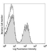
C57BL/6 mouse splenocytes were stained with FITC anti-mouse CD19 and with Alexa Fluor® 647 anti-mouse CD3ε (clone KT3.1.1, left) or rat IgG2a, κ isotype control (right). -

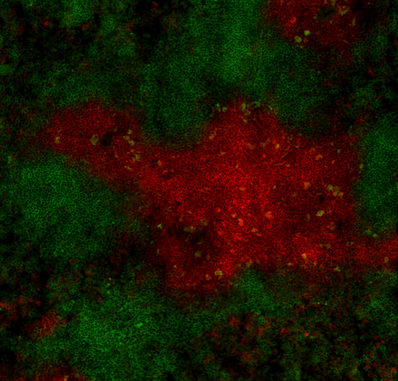
C57BL/6 mouse frozen spleen section was fixed with 4% paraformaldehyde (PFA) for 10 minutes at room temperature and blocked with 5% FBS for 30 minutes at room temperature. Then the section was stained with 10 µg/ml of anti-mouse CD3ε (clone KT3.1.1) Alexa Fluor® 647 (red), anti-mouse B220 (clone RA3-6B2) Alexa Fluor® 488 (green) overnight at 4°C. The image was captured by 10X objective.
| Cat # | Size | Price | Quantity Check Availability | Save | ||
|---|---|---|---|---|---|---|
| 155609 | 25 µg | 81€ | ||||
| 155610 | 100 µg | 184€ | ||||
CD3ε is a 20 kD transmembrane protein, also known as CD3 or T3. It is a member of the Ig superfamily and primarily expressed on T cells, NK-T cells, and at different levels on thymocytes during T cell differentiation. CD3ε forms a TCR complex by associating with the CD3δ, γ and ζ chains, as well as the TCR α/β or γ/δ chains. CD3 plays a critical role in TCR signal transduction, T cell activation, and antigen recognition by binding the peptide/MHC antigen complex.
Product DetailsProduct Details
- Verified Reactivity
- Mouse
- Antibody Type
- Monoclonal
- Host Species
- Rat
- Immunogen
- CBAT6 Thymocytes
- Formulation
- Phosphate-buffered solution, pH 7.2, containing 0.09% sodium azide.
- Preparation
- The antibody was purified by affinity chromatography and conjugated with Alexa Fluor® 647 under optimal conditions.
- Concentration
- 0.5 mg/ml
- Storage & Handling
- The antibody solution should be stored undiluted between 2°C and 8°C, and protected from prolonged exposure to light. Do not freeze.
- Application
-
FC - Quality tested
IHC-F - Verified - Recommended Usage
-
Each lot of this antibody is quality control tested by immunofluorescent staining with flow cytometric analysis. For flow cytometric staining, the suggested use of this reagent is ≤ 0.25 µg per million cells in 100 µl volume. For immunohistochemistry on frozen tissue sections, a concentration range of 5.0 - 10 µg/ml is suggested. It is recommended that the reagent be titrated for optimal performance for each application.
* Alexa Fluor® 647 has a maximum emission of 668 nm when it is excited at 633 nm / 635 nm.
Alexa Fluor® and Pacific Blue™ are trademarks of Life Technologies Corporation.
View full statement regarding label licenses - Excitation Laser
-
Red Laser (633 nm)
-
Application References
(PubMed link indicates BioLegend citation) -
- Tomonari K. 1988. Immunogenetics. 28:455. (FC)
- Product Citations
-
- RRID
-
AB_2750435 (BioLegend Cat. No. 155609)
AB_2750436 (BioLegend Cat. No. 155610)
Antigen Details
- Structure
- Ig superfamily, forms CD3/TCR complex with CD3δ, γ and ζ subunits and TCR (α/β and γ/δ), 20 kD.
- Distribution
-
Thymocytes (differentiation dependent), mature T cells, NK-T cells
- Function
- TCR signal transduction, T cell activation
- Ligand/Receptor
- TCR
- Cell Type
- NKT cells, T cells, Thymocytes, Tregs
- Biology Area
- Immunology
- Molecular Family
- CD Molecules, TCRs
- Antigen References
-
- Preston S, et al. 1997. Eur. J. Immunol. 27:1911.
- Davis MM. 1990. Annu. Rev. Biochem. 59:475.
- Weiss A, Littman DR. 1994. Cell. 76:263.
- Gene ID
- 12501 View all products for this Gene ID
- UniProt
- View information about CD3epsilon on UniProt.org
Related Pages & Pathways
Pathways


Related FAQs
Other Formats
View All CD3ε Reagents Request Custom Conjugation| Description | Clone | Applications |
|---|---|---|
| Purified anti-mouse CD3ε | KT3.1.1 | FC,IHC-F |
| FITC anti-mouse CD3ε | KT3.1.1 | FC |
| PE anti-mouse CD3ε | KT3.1.1 | FC |
| APC anti-mouse CD3ε | KT3.1.1 | FC |
| Pacific Blue™ anti-mouse CD3ε | KT3.1.1 | FC |
| Alexa Fluor® 647 anti-mouse CD3ε | KT3.1.1 | FC,IHC-F |
| APC/Fire™ 750 anti-mouse CD3ε | KT3.1.1 | FC |
| PerCP/Cyanine5.5 anti-mouse CD3ε | KT3.1.1 | FC |
| Brilliant Violet 421™ anti-mouse CD3ε | KT3.1.1 | FC |
| PE/Dazzle™ 594 anti-mouse CD3ε | KT3.1.1 | FC |
| PE/Cyanine7 anti-mouse CD3ε | KT3.1.1 | FC |
| Spark Red™ 718 anti-mouse CD3ε (Flexi-Fluor™) | KT3.1.1 | FC |
Customers Also Purchased
Compare Data Across All Formats
This data display is provided for general comparisons between formats.
Your actual data may vary due to variations in samples, target cells, instruments and their settings, staining conditions, and other factors.
If you need assistance with selecting the best format contact our expert technical support team.
-
Purified anti-mouse CD3ε

C57 splenocytes were stained with purified CD3ε (clone KT3.1... 
C57BL/6 mouse frozen spleen section was fixed with 4% parafo... -
FITC anti-mouse CD3ε

C57BL/6 mouse splenocytes were stained with CD3ε (cl... -
PE anti-mouse CD3ε

C57BL/6 mouse splenocytes were stained with FITC anti-mouse ... -
APC anti-mouse CD3ε

C57BL/6 mouse splenocytes were stained with FITC anti-mouse ... -
Pacific Blue™ anti-mouse CD3ε

C57BL/6 mouse splenocytes were stained with FITC anti-mouse ... -
Alexa Fluor® 647 anti-mouse CD3ε

C57BL/6 mouse splenocytes were stained with FITC anti-mouse ... 
C57BL/6 mouse frozen spleen section was fixed with 4% parafo... -
APC/Fire™ 750 anti-mouse CD3ε

C57BL/6 mouse splenocytes were stained with CD45R/B220 FITC ... -
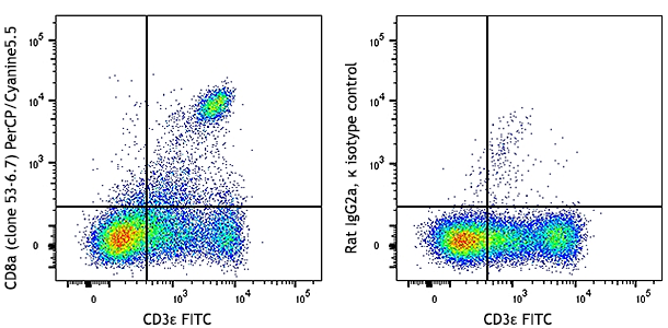
PerCP/Cyanine5.5 anti-mouse CD3ε

C57BL/6 mouse splenocytes were stained with CD45R/B220 FITC ... -
Brilliant Violet 421™ anti-mouse CD3ε

C57BL/6 mouse splenocytes were stained with APC anti-mouse C... -
PE/Dazzle™ 594 anti-mouse CD3ε

C57BL/6 mouse splenocytes were stained with APC anti-mouse C... -
PE/Cyanine7 anti-mouse CD3ε

C57BL/6 mouse splenocytes were stained with CD45R/B220 FITC ... -
Spark Red™ 718 anti-mouse CD3ε (Flexi-Fluor™)


Follow Us