 Login / Register


- Clone
- LCK-01 (See other available formats)
- Regulatory Status
- RUO
- Other Names
- Proto-oncogene tyrosine kinase lck, p56-Lck, T-cell specific protein tyrosine kinase
- Isotype
- Mouse IgG2a, κ
- Ave. Rating
- Submit a Review
- Product Citations
- publications

-

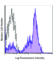
Human peripheral blood lymphocytes were surface stained with CD3 FITC, then treated with the Nuclear Factor Fixation and Permeabilization Buffer Set and intracellularly stained with anti-Lck (clone LCK-01) Alexa Fluor® 647 (top) or mouse IgG1, κ Alexa Fluor® 647 (bottom). -

| Cat # | Size | Price | Quantity Check Availability | Save | ||
|---|---|---|---|---|---|---|
| 628303 | 25 µg | 118€ | ||||
| 628304 | 100 µg | 278€ | ||||
Lck is a 56 kD tyrosine protein kinase and a member of the src subfamily that contains both an SH2 and SH3 domain. Alternatively spliced isoforms of Lck have been reported. Lck is a cytoplasmic protein bound to CD4 and CD8 in T lymphocytes that participates in antigen-induced T cell activation through phosphorylation of the T cell receptor zeta chain. Lck is activated upon T cell receptor engagement and is involved in the onset of cell cycle progression induced by interleukin-2. Phosphorylation by Csk downregulates the activity of Lck. In addition to CD4 and CD8, Lck has been reported to interact with PI3K through its SH3 domain and tyrosine phosphorylated KHDRBS1/p70 through its SH2 domain. In addition, Lck has been shown to associate with the SAP transmembrane tyrosine phosphatase. The Lck-01 monoclonal antibody recognizes human Lck and has been shown to be useful for Western blotting, immunoprecipitation, immunocytochemistry, and flow cytometry.
Product DetailsProduct Details
- Verified Reactivity
- Human
- Antibody Type
- Monoclonal
- Host Species
- Mouse
- Immunogen
- Peptide corresponding to a.a. 22-36 in human Lck
- Formulation
- Phosphate-buffered solution, pH 7.2, containing 0.09% sodium azide.
- Preparation
- The antibody was purified by affinity chromatography and conjugated with Alexa Fluor® 647 under optimal conditions.
- Concentration
- 0.5 mg/ml
- Storage & Handling
- The antibody solution should be stored undiluted between 2°C and 8°C, and protected from prolonged exposure to light. Do not freeze.
- Application
-
ICFC - Quality tested
FC - Verified - Recommended Usage
-
Each lot of this antibody is quality control tested by intracellular immunofluorescent staining using our nuclear factor staining protocol. For flow cytometric staining, the suggested use of this reagent is ≤ 0.25 µg per million cells in 100 µl volume. It is recommended that the reagent be titrated for optimal performance for each application.
* Alexa Fluor® 647 has a maximum emission of 668 nm when it is excited at 633 nm / 635 nm.
Alexa Fluor® and Pacific Blue™ are trademarks of Life Technologies Corporation.
View full statement regarding label licenses - Excitation Laser
-
Red Laser (633 nm)
- Application Notes
-
Does not cross-react with mouse.
Clone may cross-react with other Src tyrosine kinase family members. -
Application References
(PubMed link indicates BioLegend citation) -
- Romagnoli P, et al. 2001. Int. Immunol. 13:305.
- Product Citations
-
- RRID
-
AB_2564196 (BioLegend Cat. No. 628303)
AB_2564197 (BioLegend Cat. No. 628304)
Antigen Details
- Structure
- Belongs to the tyrosine family of kinases, src subfamily. Contains one SH2 domain, 1 SH3 domain. Approximately 56 kD, alternatively spliced isoforms have been reported
- Distribution
-
Cytoplasmic protein, bound to cytoplasmic CD4 and CD8 in T lymphocytes
- Function
- Participates in antigen-induced T cell activation, phosphorylates zeta chain of T cell receptor. Involved in interleukin-2 induced onset of cell cycle progression
- Interaction
- Associates with CD4 and CD8 co-receptors. Binds to PI3K through SH3 interactions, binds to tyrosine phosphorylated KHDRBS1/p70 through SH2 domain. Interacts with SAP transmembrane tyrosine phosphatase
- Modification
- Phosphorylation
- Cell Type
- T cells
- Biology Area
- Cell Biology, Signal Transduction
- Molecular Family
- Protein Kinases/Phosphatase
- Antigen References
-
1. Koga Y, et al. 1986. Eur. J. Immunol. 16:1643.
2. Vogel LB, et al. 1995. Biochem. Biophys. Acta 1264:168.
3. Vogel LB, et al. 1993. Mol. Cell. Biol. 13:7408.
4. Bergaman M, et al. 1992. EMBO J. 11:2919. - Regulation
- Activated upon T cell receptor engagement. Tyrosine phosphorylation by p50 Csk downregulates catalytic activity
- Gene ID
- 3932 View all products for this Gene ID
- UniProt
- View information about Lck on UniProt.org
Related Pages & Pathways
Pathways


Related FAQs
Other Formats
View All Lck Reagents Request Custom Conjugation| Description | Clone | Applications |
|---|---|---|
| Purified anti-Lck | LCK-01 | WB,ICC,IP,FC |
| Alexa Fluor® 647 anti-Lck | LCK-01 | ICFC,FC |
Customers Also Purchased
Compare Data Across All Formats
This data display is provided for general comparisons between formats.
Your actual data may vary due to variations in samples, target cells, instruments and their settings, staining conditions, and other factors.
If you need assistance with selecting the best format contact our expert technical support team.
-
Purified anti-Lck

Total cell lysate from Jurkat cells (lane 1, 15 µg), mouse t... 
Jurkat cells were fixed with 4% PFA for ten minutes, permeab... 
Total cell lysates (15 µg total protein) from Jurkat, MOLT4,... 
Whole cell extracts (300 µg total protein) prepared from Jur... -
Alexa Fluor® 647 anti-Lck

Human peripheral blood lymphocytes were surface stained with... 


Follow Us