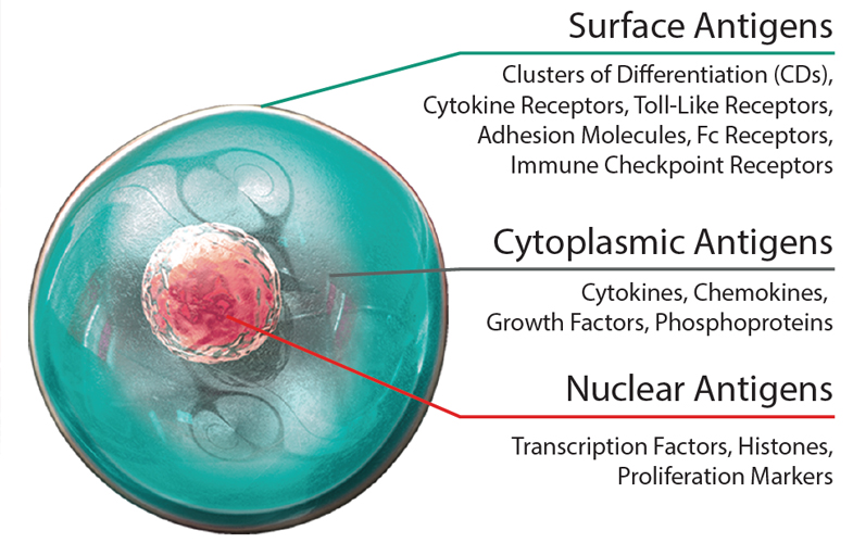
Cells often communicate with one another by secreting soluble factors that are synthesized intracellularly before being released into the external milieu. Included among these, cytokines are a diverse family of proteins that have various immunomodulatory capabilities as well as being involved in processes such as cell differentiation and directed migration. While almost all nucleated cells can generate and secrete cytokines, T helper (Th) cells and macrophages are especially productive. Cytokines exert their effects through binding to receptors expressed on the surface of target cells in an autocrine (binding to the same cell) or paracrine (binding to a nearby cell) fashion. Receptor binding triggers intracellular signaling cascades that drive gene expression.


Chemoattractant cytokines, or chemokines, induce cell movement in response to a chemical gradient (chemotaxis). Their main role is to promote the directional migration of leukocytes, including their movement from the circulatory system into tissues, and vice versa. Chemokines bind to G-protein coupled receptors (GPCRs) on the surface of target cells to initiate intracellular signaling that drives cell movement. Chemokines may also be grouped according to their function; while inflammatory chemokines recruit leukocytes in response to cytokine release by inflamed tissue, homeostatic chemokines are expressed constitutively and have an essential role in normal cellular trafficking.


Growth factors are a heterogeneous group of proteins that control the growth and differentiation of various cell types. They are often named according to their source or target, with examples including platelet-derived growth factor (PDGF), vascular endothelial growth factor (VEGF), and stem cell factor (SCF). Other intracellular proteins commonly studied in the context of immunology include transcription factors and phosphoproteins, which regulate gene expression and are involved in cellular signaling, respectively.


Intracellular Proteins

Immunoassay Quantitation of Cytokines


While various techniques can be used for measuring cytokines in biological samples such as cell culture supernatant or serum, ELISAs are often chosen for their high sensitivity. Sandwich ELISAs are especially popular as they increase specificity by using two different antibodies that each recognize a different epitope on the target molecule. We offer a broad range of products for ELISA, including off-the-shelf kits, validated antibody pairs, and a comprehensive selection of auxiliary reagents. Alternatively, our LEGENDplex™ bead-based immunoassay kits enable quantification of multiple target molecules simultaneously using a flow cytometer. LEGENDplex™ is provided as predefined panels, ranging from 3 to 14 specificities, which can additionally be mixed and matched based on researcher requirements.

Detection of Transcription Factors


Because transcription factors function by binding DNA, their analysis requires that antibody reagents used for detection are able to access the nucleus. One way of achieving this is to detect transcription factors by flow cytometry; however, the chosen staining protocol must avoid compromising cell surface markers used for identification. The True-Nuclear™ Transcription Factor Buffer Set has been specially formulated for intracellular staining with minimum effect on surface fluorophore staining and has been widely literature cited for transcription factor analysis. For those lacking access to a flow cytometer, western blots offer a quick and easy approach to detect transcription factors in cell lysates. Using high-quality antibodies that have been validated for the western blot application, it is possible to compare transcription factor expression in stimulated versus unstimulated samples, or in conditions of health versus disease.

Detection of Phosphoproteins


Phosphoprotein detection involves measuring targets that are present in the cytoplasm and is typically achieved via flow cytometry or western blot. Like transcription factor detection, phosphoprotein detection by flow cytometry must avoid damaging the cell surface markers required for identification. We have developed True-Phos™ Perm Buffer for flow cytometry, which is specifically designed to help detect our highly sensitive phosphorylation-specific antibodies. True-Phos™ Perm Buffer is not designed for use in western blotting or microscopy applications.


Accurate detection of phosphoproteins by western blot relies on the use of antibody pairs, where one antibody is specific for the phosphorylated form of a protein while the other detects total or unphosphorylated levels. We offer a wide array of phosphoprotein-specific antibodies in addition to PhosphoPair Antibody Sets that are validated for western blotting.

ProductsHere

Login / Register
Remember me
Forgot your password? Reset password?
Create an Account