 Login / Register


- Clone
- TUJ1 (See other available formats)
- Regulatory Status
- RUO
- Other Names
- CDCBM, CDCBM1, CFEOM3, CFEOM3A, FEOM3, TUBB4, Tubulin beta-3 chain, tubulin beta-III, tubulin beta-4 chain, class III beta-tubulin
- Isotype
- Mouse IgG2a, κ
- Ave. Rating
- Submit a Review
- Product Citations
- publications

-

ICC staining of Alexa Fluor® 647 anti-Tubulin β 3 (TUBB3) antibody (clone TUJ1) on SH-SY5Y neuroblastoma cells. The cells were fixed with 4% PFA, permeabilized with 0.1% Triton X-100, and blocked with 2% Normal Goat Serum. The cells were then stained with 1 µg/mL of the primary antibody for three hours at room temperature. Nuclei were counterstained with Hoechst 33342. The image was captured with a 60X objective. -

ICC staining of Alexa Fluor® 647 anti-Tubulin β 3 (TUBB3) (clone TUJ1) on SH-SY5Y neuroblastoma cells. The cells were fixed with 4% PFA, permeabilized with 0.1% Triton X-100, and blocked with 2% normal goat serum and 0.02% BSA. The cells were then stained with 1 µg/ml of the primary antibody for three hours at room temperature. Nuclei were counterstained with DAPI. The image was captured with a 40X objective. -

Paraformaldehyde-fixed (4%), 500 μm-thick mouse kidney section was processed according to the Ce3DTM Tissue Clearing Kit protocol (cat. no. 427701). The section was costained with anti-mouse Podoplanin Antibody (clone PMab-1) Alexa Fluor® 488 at 5 µg/mL (green), anti-mouse CD31 Antibody (clone MEC13.3) Alexa Fluor® 594 at 5 µg/mL (orange), and anti-Tubulin β 3 (TUBB3) Antibody (Clone TUJ1) Alexa Fluor® 647 at 5 µg/mL (magenta). The section was then optically cleared and mounted in a sample chamber. The image was captured with a 20X objective using Zeiss 780 confocal microscope and processed by Imaris image analysis software.
Watch the video. -

IHC staining of Alexa Fluor® 647 anti-Tubulin β 3 (TUBB3) (clone TUJ1) on formalin-fixed paraffin-embedded human brain tissue. Following antigen retrieval using Tris-EDTA pH 9.0, the tissue was incubated without (panel A) and with (panel B) 5.0 µg/mL of Alexa Fluor® 647 anti-Tubulin β 3 (TUBB3) (clone TUJ1) (magenta). Nuclei were counterstained with DAPI (blue) (Cat. No. 422801). Images were captured with a 10X objective. Scale bar: 50 µm
| Cat # | Size | Price | Quantity Check Availability | Save | ||
|---|---|---|---|---|---|---|
| 801209 | 25 µg | 113€ | ||||
| 801210 | 100 µg | 282€ | ||||
Tubulin is the main component of microtubules. In adults, tubulin beta 3 (TUBB3) is primarily expressed in neurons and is commonly used as a neuronal marker. It plays an important role in neuronal cell proliferation and differentiation. Mutations in this gene cause congenital fibrosis of the type 3 extraocular muscles. Tubulin beta 3 (TUBB3) is also found in a wide range of tumors. Studies indicate that it is a predictive and prognostic marker in various tumors.
Product DetailsProduct Details
- Verified Reactivity
- Human, Mouse, Rat
- Antibody Type
- Monoclonal
- Host Species
- Mouse
- Immunogen
- This antibody was raised against microtubules derived from rat brain.
- Formulation
- Phosphate-buffered solution, pH 7.2, containing 0.09% sodium azide.
- Preparation
- The antibody was purified by affinity chromatography and conjugated with Alexa Fluor® 647 under optimal conditions.
- Concentration
- 0.5 mg/ml
- Storage & Handling
- The antibody solution should be stored undiluted between 2°C and 8°C, and protected from prolonged exposure to light. Do not freeze.
- Application
-
ICC - Quality tested
ICFC, 3D IHC, IHC-P - Verified - Recommended Usage
-
Each lot of this antibody is quality control tested by immunocytochemistry. For immunocytochemistry, a concentration range of 1.0 - 5.0 μg/mL is recommended. For flow cytometric staining, the suggested use of this reagent is ≤ 5.0 µg per million cells. For 3D immunohistochemistry on formalin-fixed tissues, a concentration of 5.0 µg/mL is suggested. For immunohistochemistry on formalin-fixed paraffin-embedded tissue sections, a concentration range of 1 - 10 µg/mL is suggested. It is recommended that the reagent be titrated for optimal performance for each application.
* Alexa Fluor® 647 has a maximum emission of 668 nm when it is excited at 633 nm / 635 nm.
Alexa Fluor® and Pacific Blue™ are trademarks of Life Technologies Corporation.
View full statement regarding label licenses - Excitation Laser
-
Red Laser (633 nm)
- Application Notes
-
Additional reported applications (for the relevant formats) include: flow cytometry4, immunofluorescence microscopy1-5,7, immunohistochemistry5,7, and Western blotting8.
This antibody is well characterized and highly reactive to neuron specific Class III ß-tubulin (ßIII). TUJ1 does not identify ß-tubulin found in glial cells. TUJ1 recognizes an epitope located within the last 15 C-terminal residues8. -
Application References
(PubMed link indicates BioLegend citation) -
- Nishimura K, et al. 2017. PLoS One. 12(1): e0170568. (ICC)
- Jongbloets J, et al. 2017. Nat Commun. 8: 14666. (ICC) PubMed
- Liu W.J, et al. 2015. Eur J Histochem. 59(1): 2464. (ICC) PubMed
- Chintalapudi SR, et al. 2016. Front Aging Neursci. 8:93. (FC, ICC) PubMed
- Ambasudhan R, et al. 2011. Cell Stem Cell. 9(2):113. (IHC, ICC)
- Hu X., et al. 2006. Nature Neuroscicene. 9(12):1520. (WB) PubMed
- Zechner D., et al. 2003. Develop Biology. 258(2):406. (ICC, IHC)
- Lee MK, et al. 1990. Proc. Natl. Acad. Sci. USA 18:7195. (WB)
- Product Citations
-
- RRID
-
AB_2686930 (BioLegend Cat. No. 801209)
AB_2686931 (BioLegend Cat. No. 801210)
Antigen Details
- Structure
- Tubulin β 3 is a 450 amino acid protein with a molecular mass of ~50 kD.
- Distribution
-
Tissue distribution: central and peripheral nervous system.
Cellular distribution: cytosol, cytoskeleton and nucleus. - Function
- Tubulin β 3 is the major constituent of microtubules, and plays a critical role in proper axon guidance and maintenance.
- Interaction
- Alpha tubulin, kinesin and dynein.
- Cell Type
- Mature Neurons, Neural Stem Cells
- Biology Area
- Cell Biology, Neuroscience, Neuroscience Cell Markers, Stem Cells
- Molecular Family
- Microtubules
- Antigen References
-
1. Zhao X, et al. 2017. Med Sci Monit. 22: 3915.
2. Lebok P, et al. 2016. Oncol Lett. 11(3):1987.
3. Du J, et al. 2015. BMC Cancer. 15:536. PubMed
4. Rogue DM., et al. 2013. Clin Exp Metastasis. 31(1): 101.
5. Ploussard G, et al. 2010. Cancer Res. 70(22):9253. PubMed - Gene ID
- 10381 View all products for this Gene ID
- Specificity (DOES NOT SHOW ON TDS):
- Tubulin beta-3
- Specificity Alt (DOES NOT SHOW ON TDS):
- Tubulin β 3 (TUBB3)
- App Abbreviation (DOES NOT SHOW ON TDS):
- ICC,ICFC,3D IHC,IHC-P
- UniProt
- View information about Tubulin beta-3 on UniProt.org


Related FAQs
Other Formats
View All Tubulin β 3 (TUBB3) Reagents Request Custom Conjugation| Description | Clone | Applications |
|---|---|---|
| Alexa Fluor® 488 anti-Tubulin β 3 (TUBB3) | TUJ1 | ICC,IHC-P,ICFC,3D IHC,SB |
| Purified anti-Tubulin β 3 (TUBB3) | TUJ1 | IHC-P,WB,ICC,FC,SB |
| Alexa Fluor® 594 anti-Tubulin β 3 (TUBB3) | TUJ1 | ICC,3D IHC,IHC-P |
| Alexa Fluor® 647 anti-Tubulin β 3 (TUBB3) | TUJ1 | ICC,ICFC,3D IHC,IHC-P |
| HRP anti-Tubulin β 3 (TUBB3) | TUJ1 | WB,IHC-P |
| Biotin anti-Tubulin β 3 (TUBB3) | TUJ1 | WB,IHC-P |
| APC anti-Tubulin β 3 (TUBB3) | TUJ1 | ICFC |
| PE/Cyanine7 anti-Tubulin β 3 (TUBB3) | TUJ1 | ICFC |
| PerCP/Cyanine5.5 anti-Tubulin β 3 (TUBB3) | TUJ1 | ICFC |
| PE anti-Tubulin β 3 (TUBB3) | TUJ1 | ICFC |
Customers Also Purchased
Compare Data Across All Formats
This data display is provided for general comparisons between formats.
Your actual data may vary due to variations in samples, target cells, instruments and their settings, staining conditions, and other factors.
If you need assistance with selecting the best format contact our expert technical support team.
-
Alexa Fluor® 488 anti-Tubulin β 3 (TUBB3)

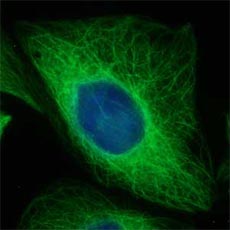
CHO cell line expressing Tubulin β 3 stained with Alexa Fluo... 
IHC staining of Alexa Fluor® 488 anti-Tubulin β 3 (TUBB3) an... 
IHC staining of Alexa Fluor® 488 anti-Tubulin β 3 (TUBB3) an... 
Human lung adenocarcinoma cell line A549 was treated with Fi... 
Paraformaldehyde-fixed (4%), 500 µm-thick mouse kidney tissu... 
Confocal image of human jejunum sample acquired using the IB... -
Purified anti-Tubulin β 3 (TUBB3)

IHC staining of purified anti-Tubulin β 3 (TUBB3) antibody (... 
IHC staining of purified anti-Tubulin β 3 (TUBB3) antibody (... 
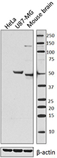
Western blot of purified anti-Tubulin β 3 (TUBB3) antibody (... 
ICC staining of purified anti-Tubulin β 3 (TUBB3) antibody (... 
Methanol fixed superior cervical ganglion neurons were stain... -
Alexa Fluor® 594 anti-Tubulin β 3 (TUBB3)

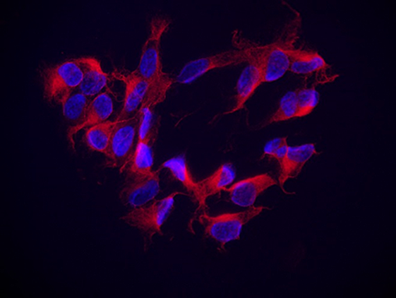
ICC staining of Alexa Fluor® 594 anti-Tubulin β 3 (TUBB3) (c... 
Paraformaldehyde-fixed (4%), 500 μm-thick mouse kidney secti... 
A: IHC staining of Alexa Fluor® 594 anti-Tubulin β 3 (TUBB3)... -
Alexa Fluor® 647 anti-Tubulin β 3 (TUBB3)

ICC staining of Alexa Fluor® 647 anti-Tubulin β 3 (TUBB3) an... 
ICC staining of Alexa Fluor® 647 anti-Tubulin β 3 (TUBB3) (c... 
Paraformaldehyde-fixed (4%), 500 μm-thick mouse kidney secti... 
IHC staining of Alexa Fluor® 647 anti-Tubulin β 3 (TUBB3) (c... -
HRP anti-Tubulin β 3 (TUBB3)

IHC staining of HRP anti-Tubulin β 3 (TUBB3) antibody (clone... 
IHC staining of anti-Tubulin β 3 (TUBB3) antibody (clone TUJ... 
Western blot of HRP anti-Tubulin β 3 (TUBB3) antibody (clone... -
Biotin anti-Tubulin β 3 (TUBB3)

Western blot of Biotin anti-Tubulin β 3 (TUBB3) antibody (cl... 
IHC staining of Biotin anti-Tubulin β 3 (TUBB3) antibody (cl... -
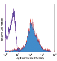
APC anti-Tubulin β 3 (TUBB3)

Human lung adenocarcinoma cell line A549 was treated with Fi... -
PE/Cyanine7 anti-Tubulin β 3 (TUBB3)

Human lung adenocarcinoma cell line A549 was treated with Fi... -
PerCP/Cyanine5.5 anti-Tubulin β 3 (TUBB3)

Human lung adenocarcinoma cell line A549 was treated with Fi... -
PE anti-Tubulin β 3 (TUBB3)

Human lung adenocarcinoma cell line A549 was treated with Fi...


Follow Us