 Login / Register


- Clone
- 13A3-1 (See other available formats)
- Regulatory Status
- RUO
- Other Names
- Signal transducer and activator of transcription 3, Acute-phase response factor, APRF, HIES
- Isotype
- Mouse IgG1, κ
- Ave. Rating
- Submit a Review
- Product Citations
- publications

-

Total cell lysates (15 µg total protein) from A431 cells serum starved and untreated (-) or stimulated with 100 ng/mL hEGF for 5 minutes were resolved by 4-12% Bis-Tris gel electrophoresis, transferred to a nitrocellulose membrane, and probed with 0.5 µg/mL (1:1000 dilution) of Purified anti-STAT3 Phospho (Tyr705) Antibody, clone 13A3-1, for 2 hours at room temperature. Proteins were visualized by chemiluminescence detection using HRP goat anti-mouse IgG Antibody (Cat. No. 405306) at a 1:3000 dilution. Purified anti-STAT3 pan Antibody, Clone 4G4B45 (Cat. 678002), was used as a pan STAT3 loading control at 1.0 µg/mL (1:500 dilution). Lane M: Molecular Weight marker. -

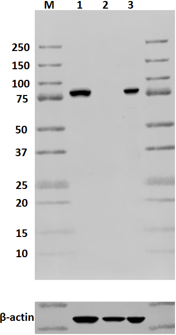
Total cell lysate from PC3 (lane 1, 15 µg), untreated HepG2 (lane 2, 15 µg), HepG2 treated with 10 ng/ml of IL-6 for 15 minutes (lane 3, 15 µg) and HeLa (lane 4, 15 µg) were resolved by electrophoresis (4-12% Bis-Tris), transferred to nitrocellulose, and probed with 1 µg/ml Purified anti-STAT3 Phospho (Tyr705) antibody (clone 13A3-1). Proteins were visualized using an HRP Goat anti-mouse IgG (clone Poly4053) antibody and chemiluminescence detection. Direct-Blot™ HRP anti-β-actin antibody (clone 2F1-1) was used as a loading control. -

15 µg of total cell lysate from untreated NIH3T3 (lane 1) and NIH3T3 treated with 100 ng/ml of IL-6 for 30 minutes (lane 2) were resolved by electrophoresis (4-20% Tris-Glycine), transferred to nitrocellulose, and probed with 1 µg/ml purified anti-STAT3 Phospho (Tyr705) antibody (clone 13A3-1). Proteins were visualized using an HRP Goat anti-mouse IgG (clone Poly4053) antibody and chemiluminescence detection. Direct-Blot™ HRP anti-β-actin antibody (clone 2F1-1) was used as a loading control. -

Serum starved A431 cells were untreated (panel A) or stimulated with 100 ng/mL hEGF for 5 minutes (panel B). Cells were fixed with 4% paraformaldehyde for 10 minutes, permeabilized with ice-cold methanol, and blocked with 5% FBS for 60 minutes. Fixed cells were then intracellularly stained with 5 µg/mL (1:100 dilution) of Purified anti-STAT3 Phospho (Tyr705) Antibody, clone 13A3-1 for 2 hours at room temperature, after which proteins were visualized with Alexa Fluor® 594 goat anti-mouse IgG (Cat. No. 405326) at 2.5 µg/mL (1:200 dilution). Nuclei were counterstained with DAPI, and the image was captured with a 60X objective. -

Chromatin Immunoprecipitation (ChIP) was performed using commercial Protein-G coated 96 well high-throughput ChIP assay kit by loading 3 µg of cross-linked chromatin samples from HeLa cells starved overnight and then treated with IL-6 with either A) 1:50 dilution of Go-ChIP-Grade™ Purified anti-STAT3 Phospho (Tyr705) (Clone 13A3-1), B) equal amount of Purified Mouse IgG1, κ Isotype Control Antibody (#400101), or C) competitor’s ChIP-grade Purified anti-STAT3 Phospho (Tyr705) Antibody and D) equal amount of matched Isotype Control Antibody as recommended by the manufacturer. The enriched DNA was purified and quantified by real-time qPCR using primers targeting human IRF1 gene region. The amount of immunoprecipitated DNA in each sample is represented as signal relative to the 5% of total amount of input chromatin.
| Cat # | Size | Price | Quantity Check Availability | Save | ||
|---|---|---|---|---|---|---|
| 651001 | 25 µg | 147 CHF | ||||
| 651002 | 100 µg | 323 CHF | ||||
Tyrosine phosphorylation of STAT3 at Tyr705 occurs in response to LIF, IL-6, leptin, OSM, EGF, PDGF, and HGF. It plays a key role in cell growth and apoptosis through mediating expression of a variety of genes in response to the stimuli.
Product DetailsProduct Details
- Verified Reactivity
- Human, Mouse
- Antibody Type
- Monoclonal
- Host Species
- Mouse
- Immunogen
- KLH conjugated modified synthetic peptide
- Formulation
-
This antibody is provided in phosphate-buffered solution, pH 7.2, containing 0.09% sodium azide.
Previous lots of this product may have been formulated with 0.1% or 0.05% NaN3 instead of 0.09% NaN3. For further information please contact BioLegend Technical Support or Customer Service. - Preparation
- The antibody was purified by affinity chromatography.
- Concentration
- 0.5 mg/ml
- Storage & Handling
- Upon receipt, store undiluted between 2°C and 8°C.
- Application
-
WB - Quality tested
ICC, ChIP - Verified - Recommended Usage
-
Each lot of this antibody is quality control tested by Western blotting. For Western blotting, the suggested use of this reagent is 0.5 - 1.0 µg per ml (1:500 - 1:1000 dilution). For immunocytochemistry, a concentration of 0.5 μg/ml is recommended. For ChIP applications, the suggested dilution is 1:50 by volume. It is recommended that the reagent be titrated for optimal performance for each application.
- Application Notes
-
The STAT3 Phospho (Tyr705) antibody recognizes the regulatory tyrosine phosphorylation of human STAT3 protein and has been shown to be useful for Western blotting.
During PD testing, this clone failed to stain PFA-fixed cells that had been permeabilized with Triton X-100. We highly recommend permeabilizing cells with methanol. - Product Citations
-
- RRID
-
AB_10897947 (BioLegend Cat. No. 651001)
AB_10900983 (BioLegend Cat. No. 651002)
Antigen Details
- Structure
- STAT3 is a 770 amino acid protein of 88 kD. It consists of a DNA binding domain, a SH2 domain, a regulatory tyrosine responsible for binding of SH2 domain, and a C-terminal transactivation domain.
- Distribution
-
Ubiquitous.
- Function
- STAT3 is tyrosine phosphorylated by receptor kinases in response to a variety of cytokines and growth factors. It forms homo- or heterodimer with STAT1 when tyrosine is phosphorylated, and then translocates to nucleus, acting as a transcription regulator. It is also essential for the differentiation of TH17 cells, which is involved in autoimmune diseases.
- Cell Sources
- Cytoplasm. Translocate to nucleus in response to tyrosine phosphorylation.
- Cell Type
- Embryonic Stem Cells, Neural Stem Cells
- Biology Area
- Cell Biology, Neuroscience, Neuroscience Cell Markers, Signal Transduction, Stem Cells, Synaptic Biology, Transcription Factors
- Molecular Family
- Phospho-Proteins
- Antigen References
-
1. Akira S, et al. 1994. Cell 77:63.
2. Zhang X, et al. 1995. Science 267:1990.
3. Sanchez-Margalet V, et al. 2001. Cell. Immunol. 211:30.
4. Simon A, et al. 2000. Science 290:144.
5. Hoey T, et al. 1999. Adv. Immunol. 71:145. - Regulation
- The small GTPase Rac1 binds and regulates activity of STAT3.
- Gene ID
- 6774 View all products for this Gene ID
- UniProt
- View information about STAT3 Phospho Tyr705 on UniProt.org

Related FAQs
Other Formats
View All STAT3 Phospho (Tyr705) Reagents Request Custom Conjugation| Description | Clone | Applications |
|---|---|---|
| Purified anti-STAT3 Phospho (Tyr705) | 13A3-1 | WB,ICC,ChIP |
| PE anti-STAT3 Phospho (Tyr705) | 13A3-1 | ICFC |
| Alexa Fluor® 488 anti-STAT3 Phospho (Tyr705) | 13A3-1 | ICFC |
| Brilliant Violet 421™ anti-STAT3 Phospho (Tyr705) | 13A3-1 | ICFC |
| Alexa Fluor® 647 anti-STAT3 Phospho (Tyr705) | 13A3-1 | ICFC |
| Direct-Blot™ HRP anti-STAT3 Phospho (Tyr705) | 13A3-1 | WB |
| PE/Cyanine5 anti-STAT3 Phospho (Tyr705) | 13A3-1 | ICFC |
| FITC anti-STAT3 Phospho (Tyr705) | 13A3-1 | ICFC |
| PerCP/Cyanine5.5 anti-STAT3 Phospho (Tyr705) | 13A3-1 | ICFC |
Customers Also Purchased
Compare Data Across All Formats
This data display is provided for general comparisons between formats.
Your actual data may vary due to variations in samples, target cells, instruments and their settings, staining conditions, and other factors.
If you need assistance with selecting the best format contact our expert technical support team.
-
Purified anti-STAT3 Phospho (Tyr705)

Total cell lysate from PC3 (lane 1, 15 µg), untreated HepG2 ... 
15 µg of total cell lysate from untreated NIH3T3 (lane 1) an... 
Total cell lysates (15 µg total protein) from A431 cells ser... 
Serum starved A431 cells were untreated (panel A) or stimula... 
Chromatin Immunoprecipitation (ChIP) was performed using com... -
PE anti-STAT3 Phospho (Tyr705)

Human whole blood was stimulated with (top), or without (bot... 
-
Alexa Fluor® 488 anti-STAT3 Phospho (Tyr705)

Human whole blood was stimulated with (top) or without (bott... 
-
Brilliant Violet 421™ anti-STAT3 Phospho (Tyr705)

Human whole blood was stimulated with (top) or without (bott... 
-
Alexa Fluor® 647 anti-STAT3 Phospho (Tyr705)

Human whole blood was stimulated with (top), or without (bot... 
-
Direct-Blot™ HRP anti-STAT3 Phospho (Tyr705)

15 µg of total protein extract from HepG2 cells untreated o... -
PE/Cyanine5 anti-STAT3 Phospho (Tyr705)

Human whole blood was stimulated with (left), or without (ri... -
FITC anti-STAT3 Phospho (Tyr705)

Human whole blood was stimulated with (left) or without (rig... -
PerCP/Cyanine5.5 anti-STAT3 Phospho (Tyr705)

Human whole blood was stimulated with (left) or without (rig...


Follow Us