 Login / Register


- Clone
- Poly19013 (See other available formats)
- Regulatory Status
- RUO
- Other Names
- Paired box protein Pax-6, oculorhombin, aniridia type II protein, Sey, Protein eyeless, PRB-278P-100
- Previously
-
Covance Catalog# PRB-278P
- Isotype
- Rabbit Polyclonal IgG
- Ave. Rating
- Submit a Review
- Product Citations
- publications

-

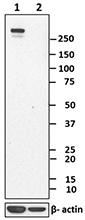
Whole cell extracts (15µg total protein) from 293T (human), (left panel) or E17 Ms. brain (mouse brain at day 17 of development), (right panel) were resolved by 4-12% Bis-Tris gel electrophoresis, transferred to PVDF membrane and probed with 1.0 µg/mL (1:2000 dilution) of purified Pax-6 rabbit antibody (clone Poly19013) for 2 hr at RT. Proteins were visualized by chemiluminescence detection using HRP Donkey anti-rabbit IgG Antibody (Cat. No. 406401, 1:3000 dilution). Direct-Blot™ HRP anti-GAPDH antibody (Cat. No. 607904) was used as a loading control at 1:25000 dilution (lower). Lane M:Molecular weight marker. -

Whole cell extracts (15 µg total protein) from either untransfected 293E cells or transfected with GFP were resolved by 4-12% Bis-Tris gel electrophoresis, transferred to PVDF membrane and probed with 1.0 µg/mL (1:2000 dilution) of purified Pax-6 rabbit antibody (clone Poly19013) (panel A) or purified anti-GFP antibody (clone 1GFP63, Cat. No. 668206) for 2 hr at RT. Proteins were visualized by chemiluminescence detection using HRP Donkey anti-rabbit IgG Antibody (Cat. No. 406401, 1:3000 dilution) or HRP Goat anti-mouse IgG Antibody (Cat. No. 405306, 1:3000 dilution). Direct-Blot™ HRP anti-GAPDH antibody (Cat. No. 607904) was used as a loading control at 1:25000 dilution (lower).Lane M:Molecular weight marker. -

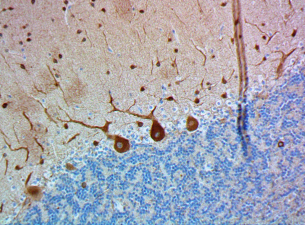
IHC staining of purified anti-Pax-6 antibody (clone Poly19013) on formalin-fixed paraffin-embedded mouse brain tissue. Following antigen retrieval using Sodium Citrate H.I.E.R., the tissue was incubated with 20 µg/ml of the primary antibody for 60 minutes at room temperature. BioLegend's Ultra-Streptavidin (USA) HRP kit (Multi-Species, DAB, Cat. No. 929901) was used for detection followed by hematoxylin counterstaining, according to the protocol provided. The image was captured with a 40X objective.
| Cat # | Size | Price | Quantity Check Availability | Save | ||
|---|---|---|---|---|---|---|
| 901302 | 25 µL | 136 CHF | ||||
| 901301 | 100 µL | 413 CHF | ||||
Pax6 is a transcription factor present during embryonic development. The encoded protein contains two different binding sites that are known to bind DNA and function as a regulator of gene transcription. It is a key regulatory gene of eye and brain development. Within the brain, the protein is involved in development of specialized cells that process smell. Pax-6 acts as a critical gene for the development of eyes and other sensory organs, certain neural and epidermal tissues as well as other homologous structures, usually derived from ectodermal tissues. Pax6 serves as a regulator in the coordination and pattern formation required for differentiation and proliferation to successfully take place, ensuring that the processes of neurogenesis and oculogenesis are carried out successfully. As a transcription factor, Pax6 acts at the molecular level in the signaling and formation of the central nervous system. The characteristic paired DNA binding domain of Pax6 utilizes two DNA-binding domains, the paired domain (PD), and the paired-type homeodomain (HD). These domains function separately. An example of this lies in HD’s regulatory involvement in the formation of the lens and retina throughout oculogenesis contrasted by the molecular mechanisms of control exhibited on the patterns of neurogenesis in brain development by PD. The HD and PD domains act in close coordination, giving Pax6 its multifunctional nature in directing molecular signaling in formation of the CNS.
The vertebrate PAX6 locus encodes at least three different protein isoforms, these being the canonical PAX6, PAX6(5a), and PAX6(ΔPD). The canonical PAX6 protein contains an N-terminal paired domain, connected by a linker region to a paired-type homeodomain, and a proline/serine/threonine (P/S/T)-rich C-terminal domain. The paired domain and paired-type homeodomain each have DNA binding activities, while the P/S/T-rich domain possesses a transactivation function. PAX6(5a) is a product of the alternatively spliced exon 5a resulting in a 14 residue insertion in the paired domain which alters the specificity of this DNA binding activity. The nucleotide sequence corresponding to the linker region encodes a set of three alternative translation start codons from which the third PAX6 isoform originates. Collectively known as the PAX6(ΔPD) or pairedless isoforms, these three gene products all lack a paired domain. The pairedless proteins possess molecular weights of 43, 33, or 32kDa, depending on the particular start codon used. PAX6 transactivation function is attributed to the variable length C-terminal P/S/T-rich domain which stretches to 153 residues in human and mouse proteins.
Product Details
- Verified Reactivity
- Human, Mouse, Rat
- Antibody Type
- Polyclonal
- Host Species
- Rabbit
- Immunogen
- This antibody was generated against the peptide (QVPGSEPDMSQYWPRLQ) derived from the C-terminus of the mouse Pax-6 protein.
- Formulation
- Phosphate-buffered solution + 0.03% Thimerosal.
- Preparation
- The antibody was purified by affinity chromatography.
- Concentration
- 2 mg/mL
- Storage & Handling
- The antibody solution should be stored undiluted between 2°C and 8°C. Please note the storage condition for this antibody has been changed from -20°C to between 2°C and 8°C. You can also check your vial or your CoA to find the most accurate storage condition for this antibody.
- Application
-
WB, IHC-P - Quality tested
IHC-F - Reported in the literature, not verified in house - Recommended Usage
-
Each lot of this antibody is quality control tested by Western blotting and formalin-fixed paraffin-embedded immunohistochemical staining of brain tissue. For Western blotting, the suggested use of this reagent is 1.0 µg/mL (1:2000). For immunohistochemistry, a dilution of 1:50 - 1:100 is suggested. It is recommended that the reagent be titrated for optimal performance for each application.
- Application Notes
-
This antibody weakly reacts with rat. It has also shown reactivity with GFP and is therefore not recommended for use in GFP-expressing systems (as determined by in-house testing).
Pax-6 has two isoforms in human and mice at 46.6 and 48.2 Kd. This antibody recognizes both. The sequence is highly conserved among Pax-6 of various species. The antibody was subsequently purified on a Protein A column and is useful in studying brain, neuronal and olfactory development in higher eukaryotes.
This clone is not recommended for ChIP (Chromatin Immunoprecipitation) assays (as determined by in-house testing). -
Application References
(PubMed link indicates BioLegend citation) -
- Tang K, et al. 2012. Development. 139:1630. (IF) PubMed
- Bandah D, et al. 2007. Invest. Ophthalmol. Vis. Sci. 48:2503. (WB) PubMed
- Marquardt T, et al. 2001. Cell. 105:43.
- Yamamoto Y, Jeffery WR. 2000. Science. 289:631
- Ashery-Padan R, et al. 2000. Genes Dev. 14:2701.
- Osumi N, et al. 1997. Development. 124:2961.
- Koroma BM, et al. 1997. Invest. Ophthalmol Vis Sci. 38:108.
- Davis JA, Reed RR. 1996. J Neurosci. 16:5082. (IHC)
- Quadrato G, et al. Nature. 545:48. (IF) PubMed
- Product Citations
-
- RRID
-
AB_2749901 (BioLegend Cat. No. 901302)
AB_2565003 (BioLegend Cat. No. 901301)
Antigen Details
- Cell Type
- Neural Stem Cells
- Biology Area
- Cell Biology, Neuroscience, Neuroscience Cell Markers, Signal Transduction, Stem Cells, Synaptic Biology, Transcription Factors
- Molecular Family
- Nuclear Markers
- Gene ID
- 5080 View all products for this Gene ID 18508 View all products for this Gene ID 25509 View all products for this Gene ID
- UniProt
- View information about Pax-6 on UniProt.org

Related FAQs
Other Formats
View All Pax-6 Reagents Request Custom Conjugation| Description | Clone | Applications |
|---|---|---|
| Purified anti-Pax-6 | Poly19013 | WB,IHC-P,IHC-F |
Customers Also Purchased
Compare Data Across All Formats
This data display is provided for general comparisons between formats.
Your actual data may vary due to variations in samples, target cells, instruments and their settings, staining conditions, and other factors.
If you need assistance with selecting the best format contact our expert technical support team.


Follow Us