 Login / Register


- Clone
- SMI 91 (See other available formats)
- Regulatory Status
- RUO
- Other Names
- CNPase, Cnp-1, Cnp1, 2',3'-cyclic-nucleotide 3'-phosphodiesterase
- Isotype
- Mouse IgG1, κ
- Ave. Rating
- Submit a Review
- Product Citations
- publications

-

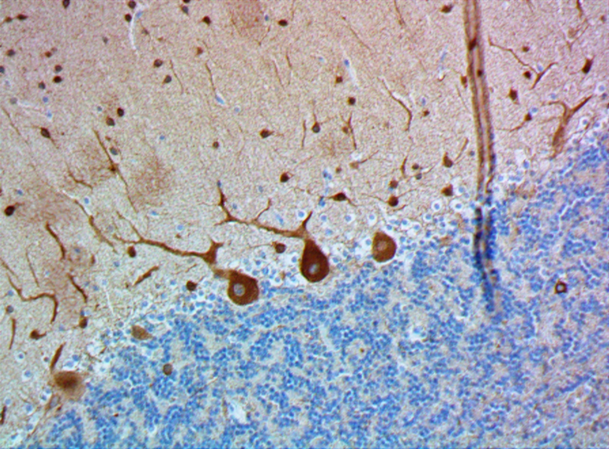
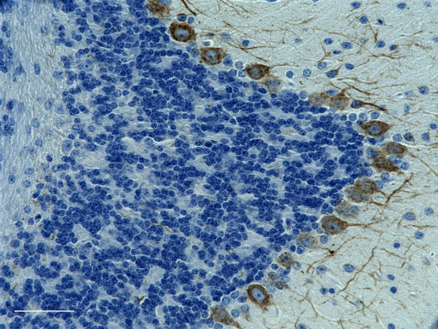
IHC staining of purified anti-Myelin CNPase antibody (clone SMI 91) on formalin-fixed paraffin-embedded mouse brain tissue. Following antigen retrieval using Retrieve-All Antigen Unmasking System 3: Acidic, 10X (Cat. No. 927801), the tissue was incubated with 1 µg/ml of the primary antibody for 60 minutes at room temperature. BioLegend´s Ultra-Streptavidin (USA) HRP kit (Multi-Species, DAB, Cat. No. 929901) was used for detection followed by hematoxylin counterstaining, according to the protocol provided. The image was captured with a 40X objective. Scale bar: 50 µm -

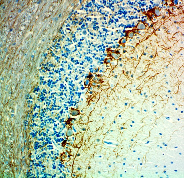
IHC staining of purified anti-Myelin CNPase antibody (clone SMI 91) on formalin-fixed paraffin-embedded rat brain tissue. Following antigen retrieval using Retrieve-All Antigen Unmasking System 3: Acidic, 10X (Cat. No. 927801), the tissue was incubated with 1 µg/ml of the primary antibody for 60 minutes at room temperature. BioLegend´s Ultra-Streptavidin (USA) HRP kit (Multi-Species, DAB, Cat. No. 929901) was used for detection followed by hematoxylin counterstaining, according to the protocol provided. The image was captured with a 40X objective. Scale bar: 50 µm -

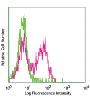
Western blot of purified anti-Myelin CNPase antibody (clone SMI 91). Lane 1: Molecular weight marker; Lane 2: 30 µg of human brain lysate; Lane 3: 30 µg of mouse brain lysate; Lane 4: 30 µg of rat brain lysate. The blot was incubated with 1 µg/mL of the primary antibody overnight at 4°C, followed by incubation with HRP labeled goat anti-mouse IgG (Cat. No. 405306). Enhanced chemiluminescence was used as the detection system.
| Cat # | Size | Price | Quantity Check Availability | Save | ||
|---|---|---|---|---|---|---|
| 836403 | 25 µg | 112 CHF | ||||
| 836404 | 100 µg | 276 CHF | ||||
High CNPase expression is seen in myelin producing cells, including oligodendrocytes and Schwann cells. CNPase accounts for roughly 4% of the total myelin protein in the central nervous system (CNS). CNPase binds to tubulin heterodimers and plays a role in tubulin polymerization, and oligodendrocyte process outgrowth. The enzyme isolated from the mammalian brain is primarily a mixed dimer of approximately 94 kD. The dimer consists of a varied proportion of CNP1 (46 kD) and CNP2 (48 kD) subunits in various species. Since the enzyme is a myelin-associated enzyme, it is of considerable interest in the study of diseases and disorders in which myelin is affected, such as multiple sclerosis, subacute sclerosing panencephalitis, acquired immunodeficiency with CNS involvement, and peripheral neuropathies. The combination of clone SMI 91 with clone SMI 94 and/or clone SMI 99 is useful for immunocytochemical studies on the progression of normal and pathologic myelination.
Product DetailsProduct Details
- Verified Reactivity
- Human, Rat, Mouse
- Antibody Type
- Monoclonal
- Host Species
- Mouse
- Formulation
- Phosphate-buffered solution, pH 7.2, containing 0.09% sodium azide.
- Preparation
- The antibody was purified by affinity chromatography.
- Concentration
- 0.5 mg/ml
- Storage & Handling
- The antibody solution should be stored undiluted between 2°C and 8°C.
- Application
-
IHC-P - Quality tested
WB - Verified
IHC-F, ICC, EM, IP - Reported in the literature, not verified in house - Recommended Usage
-
Each lot of this antibody is quality control tested by formalin-fixed paraffin-embedded immunohistochemical staining. For immunohistochemistry, a concentration of 1.0 µg/mL. For Western blotting, the suggested use of this reagent is 0.5 - 5.0 µg/mL. It is recommended that the reagent be titrated for optimal performance for each application.
- Application Notes
-
Additional reported applications (for the relevant formats) include: immunohistochemical staining on frozen tissue sections1,2,4,8 (IHC), immunocytochemistry5,6 (ICC), and western blot (WB).3,7
-
Application References
(PubMed link indicates BioLegend citation) -
- Samanta J, et al. 2010. Stroke. 2:357-62. (IHC-F)
- Werner HB, et al. 2007. J. Neurosci. 27:7717-7730. (IHC-P) PubMed
- McFerran B, et al. 1997. J Cell Sci. 110:2979-2985. (WB)
- Trolle C, et al. 2014. BMC Neurosci. 15:60. (IHC-F) PubMed
- Liyanage VR, et al. 2013. Mol. Autism 4:46. (ICC)
- Göttle P, et al. 2015. J. Neurosci. 35:906. (ICC)
- Yu C, et al. 2008. Glia. 56:877. (WB)
- Johnson GC, et al. 2014. Pathol. 51:146. (IHC-P) PubMed
- Product Citations
-
- RRID
-
AB_2728547 (BioLegend Cat. No. 836403)
AB_2566639 (BioLegend Cat. No. 836404)
Antigen Details
- Structure
- Reacts with the 46 kD and 48 kD subunits of the 94 kD myelin CNPase dimer.
- Distribution
-
Tissue distribution: Central and peripheral nervous systems.
Cellular distribution: Cytoskeleton, nucleus, mitochondria, cytosol, and plasma membrane. - Function
- CNPase detects developing and adult myelin, and developing oligodendrocytes and Schwann cells. CNPase distinguishes oligodendrocytes from astrocytes, microglia, neurons, and other cells in brain sections.
- Interaction
- Detects developing and adult myelin and developing oligodendrocytes and Schwann cells. Distinguishes oligodendrocytes from astrocytes, microglia, neurons, and other cells in brain sections.
- Cell Type
- Oligodendrocytes
- Biology Area
- Cell Biology, Neuroscience, Neuroscience Cell Markers
- Molecular Family
- Enzymes and Regulators, Phospho-Proteins
- Antigen References
-
- Han H, et al. 2013. Biofactors. 39(3):233.
- Raasakka A, et al. 2014. Neurosci Bull. 30(6):956.
- Kim JY, et al. 2003. J. Neurosci. 23(13):5561-5571.
- Sprinkle TJ, et al. 1989. Crit. Rev. Neurobiol. 4:235-301.
- Ding BS, et al. 2013. PLoS One 8:e62150.
- Ferletta M, et al. 2011. Int. J. Cancer 129:45.
- Gene ID
- 1267 View all products for this Gene ID
- UniProt
- View information about Myelin CNPase on UniProt.org

Related FAQs
Other Formats
View All Myelin CNPase Reagents Request Custom Conjugation| Description | Clone | Applications |
|---|---|---|
| Purified anti-Myelin CNPase | SMI 91 | IHC-P,WB |
| Alexa Fluor® 647 anti-Myelin CNPase | SMI 91 | IHC-P |
Customers Also Purchased
Compare Data Across All Formats
This data display is provided for general comparisons between formats.
Your actual data may vary due to variations in samples, target cells, instruments and their settings, staining conditions, and other factors.
If you need assistance with selecting the best format contact our expert technical support team.
-
Purified anti-Myelin CNPase

IHC staining of purified anti-Myelin CNPase antibody (clone ... 
IHC staining of purified anti-Myelin CNPase antibody (clone ... 
Western blot of purified anti-Myelin CNPase antibody (clone ... -
Alexa Fluor® 647 anti-Myelin CNPase

IHC staining of Alexa Fluor® 647 anti-Myelin CNPase antibody... 
IHC staining of Alexa Fluor® 647 anti-Myelin CNPase antibody... 
IHC staining of Alexa Fluor® 647 anti-Myelin CNPase antibody... 
IHC staining of Alexa Fluor® 647 anti-Myelin CNPase antibody...


Follow Us