 Login / Register


- Clone
- Poly9060 (See other available formats)
- Regulatory Status
- RUO
- Other Names
- Keratin, type I cytoskeletal 14, CK-14, cytokeratin 14, keratin 14 (epidermolysis bullosa simplex, Dowling-Meara, Koebner), K14, CK14
- Isotype
- Chicken IgY
- Ave. Rating
- Submit a Review
- Product Citations
- publications

-

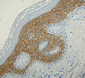
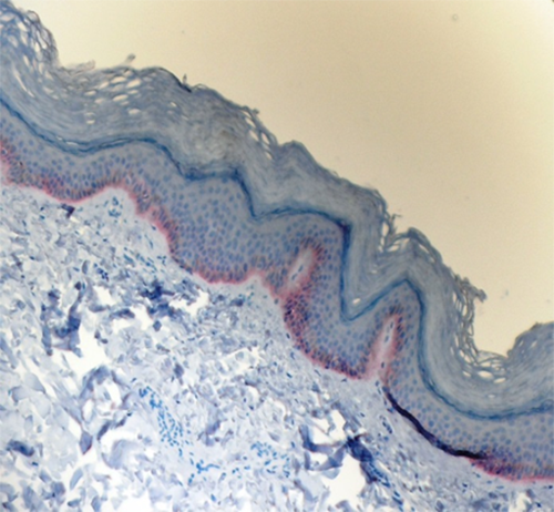
IHC staining of purified anti-Keratin 14 antibody (Poly9060) on formalin-fixed paraffin-embedded canine skin tissue. Following antigen retrieval using Sodium Citrate H.I.E.R., the tissue was incubated with 1 µg/mL of the primary antibody for 60 minutes at room temperature. After incubation with biotinylated anti-chicken antibody, the tissue was labeled with BioLegend's HRP labeling reagent followed by addition of AEC chromogen concentrate (Cat. No. 925804). The tissue was then counterstained with hematoxylin. The image was captured with a 40X objective. Scale bar: 50 µm -

IHC staining of purified anti-Keratin 14 antibody (Poly9060) on formalin-fixed paraffin-embedded primate skin tissue. Following antigen retrieval using Sodium Citrate H.I.E.R., the tissue was incubated with 1 µg/mL of the primary antibody for 60 minutes at room temperature. After incubation with biotinylated anti-chicken antibody, the tissue was labeled with BioLegend's HRP labeling reagent followed by addition of AEC chromogen concentrate (Cat. No. 925804). The tissue was then counterstained with hematoxylin. The image was captured with a 40X objective. Scale bar: 50 µm -

IHC staining of purified anti-Keratin 14 antibody (Poly9060) on formalin-fixed paraffin-embedded mouse skin tissue. Following antigen retrieval using Sodium Citrate H.I.E.R., the tissue was incubated with 1 µg/mL of the primary antibody for 60 minutes at room temperature. After incubation with biotinylated anti-chicken antibody, the tissue was labeled with BioLegend's HRP labeling reagent followed by addition of AEC chromogen concentrate (Cat. No. 925804). The tissue was then counterstained with hematoxylin. The image was captured with a 40X objective. Scale bar: 50 µm -

IHC staining of purified anti-Keratin 14 antibody (Poly9060) on formalin-fixed paraffin-embedded rat skin tissue. Following antigen retrieval using Sodium Citrate H.I.E.R., the tissue was incubated with 1 µg/mL of the primary antibody for 60 minutes at room temperature. After incubation with biotinylated anti-chicken antibody, the tissue was labeled with BioLegend’s HRP labeling reagent followed by addition of AEC chromogen concentrate (Cat. No. 925804). The tissue was then counterstained with hematoxylin. The image was captured with a 40X objective. Scale bar: 50 µm -

Western blot analysis of cell lysates from HaCaT (positive control), THP-1 (low expression negative control) and UMR106 (Rat) cells using Keratin 14 Chicken primary antibody (clone Poly19060, 1:500 dilution) and HRP Donkey anti-chicken secondary antibody (1:3000 dilution). Direct-Blot™ HRP anti-β-actin (Cat. No. 643807) was used as a loading control (1:8000 dilution). -

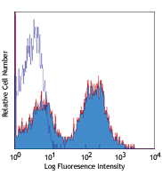
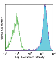
HaCaT cells were fixed with 4% paraformaldehyde (PFA) for 15 minutes, permeabilized with 0.5% Triton X-100 for 3 minutes, and blocked with 5% FBS for 60 minutes. Then the cells were intracellularly stained with (A) Chicken IgY isotype control (Negative, Cat. No. 402101) or (B-D) Keratin 14 primary antibody (Clone Poly9060) overnight at 4°C followed by Alexa Fluor® 488 (Green) Goat anti-chicken IgY incubation for one hour at room temperature. Nuclei were counterstained with DAPI (Blue, Cat. No. 422801). The image was captured with a 60X objective using KEYENCE BZ-X700 fluorescence microscope. Exposure time (Seconds) for (A-D) is 1/30. Concentrations for (A-B) is 5 µg/ml (1:100 dilution), (C) is 2 µg/ml (1:250 dilution) and (D) is 1 µg/ml (1:500 dilution).
| Cat # | Size | Price | Quantity Check Availability | Save | ||
|---|---|---|---|---|---|---|
| 906004 | 100 µg | 350 CHF | ||||
Keratin 14 is a member of the keratin family, which is the most diverse group of intermediate filaments. Keratin 14 is a type I keratin, and is usually found as a heterotetramer with two keratin 5 molecules, a type II keratin. Together they form the cytoskeleton of epithelial cells. Mutations in the genes for these keratins are associated with epidermolysis bullosa simplex.
Product DetailsProduct Details
- Verified Reactivity
- Human, Mouse, Rat, Non-Human Primate, Dog
- Antibody Type
- Polyclonal
- Host Species
- Chicken
- Immunogen
- This monospecific polyclonal antibody was raised against a peptide sequence derived from the C-terminus of the mouse keratin 14 protein.
- Formulation
- Phosphate-buffered solution, pH 7.2, containing 0.09% sodium azide.
- Preparation
- The antibody was purified by affinity chromatography.
- Concentration
- 0.5 mg/ml
- Storage & Handling
- The antibody solution should be stored undiluted between 2°C and 8°C.
- Application
-
IHC-P - Quality tested
ICC, WB - Verified
SB - Reported in the literature, not verified in house - Recommended Usage
-
Each lot of this antibody is quality control tested by formalin-fixed paraffin-embedded immunohistochemical staining. For immunohistochemisty, a concentration of 1.0 μg/mL is suggested. For immunocytochemistry, a concentration range of 1.0 - 2.0 μg/mL is recommended. For western blotting, the suggested usage is 1.0 µg/mL. It is recommended that the reagent be titrated for optimal performance for each application.
- Application Notes
-
Additional reported applications for the relevant formats include: spatial biology (IBEX)1,2.
- Additional Product Notes
-
Iterative Bleaching Extended multi-pleXity (IBEX) is a fluorescent imaging technique capable of highly-multiplexed spatial analysis. The method relies on cyclical bleaching of panels of fluorescent antibodies in order to image and analyze many markers over multiple cycles of staining, imaging, and, bleaching. It is a community-developed open-access method developed by the Center for Advanced Tissue Imaging (CAT-I) in the National Institute of Allergy and Infectious Diseases (NIAID, NIH).
-
Application References
(PubMed link indicates BioLegend citation) - Product Citations
-
- RRID
-
AB_2616962 (BioLegend Cat. No. 906004)
Antigen Details
- Structure
- Keratin 14 is a 52 kD protein.
- Distribution
-
Cytoplasm, extracellular, and nucleus.
- Function
- Nonhelical tail domain is involved in promoting KRT5-KRT14 filaments to self-organize into large bundles, enhances the mechanical properties involved in resilience of keratin intermediate filaments in vitro.
- Biology Area
- Cell Biology, Cell Motility/Cytoskeleton/Structure, Neuroscience, Neuroscience Cell Markers
- Molecular Family
- Intermediate Filaments
- Antigen References
-
- Yuspa SH, et al. 1989. J. Cell Biol. 109:1207.
- Roop RD, et al. 1984. J. Biol. Chem. 259:8037.
- Gene ID
- 3861 View all products for this Gene ID
- UniProt
- View information about Keratin 14 on UniProt.org

Related FAQs
- If an antibody clone has been previously successfully used in IBEX in one fluorescent format, will other antibody formats work as well?
-
It’s likely that other fluorophore conjugates to the same antibody clone will also be compatible with IBEX using the same sample fixation procedure. Ultimately a directly conjugated antibody’s utility in fluorescent imaging and IBEX may be specific to the sample and microscope being used in the experiment. Some antibody clone conjugates may perform better than others due to performance differences in non-specific binding, fluorophore brightness, and other biochemical properties unique to that conjugate.
- Will antibodies my lab is already using for fluorescent or chromogenic IHC work in IBEX?
-
Fundamentally, IBEX as a technique that works much in the same way as single antibody panels or single marker IF/IHC. If you’re already successfully using an antibody clone on a sample of interest, it is likely that clone will have utility in IBEX. It is expected some optimization and testing of different antibody fluorophore conjugates will be required to find a suitable format; however, legacy microscopy techniques like chromogenic IHC on fixed or frozen tissue is an excellent place to start looking for useful antibodies.
- Are other fluorophores compatible with IBEX?
-
Over 18 fluorescent formats have been screened for use in IBEX, however, it is likely that other fluorophores are able to be rapidly bleached in IBEX. If a fluorophore format is already suitable for your imaging platform it can be tested for compatibility in IBEX.
- The same antibody works in one tissue type but not another. What is happening?
-
Differences in tissue properties may impact both the ability of an antibody to bind its target specifically and impact the ability of a specific fluorophore conjugate to overcome the background fluorescent signal in a given tissue. Secondary stains, as well as testing multiple fluorescent conjugates of the same clone, may help to troubleshoot challenging targets or tissues. Using a reference control tissue may also give confidence in the specificity of your staining.
- How can I be sure the staining I’m seeing in my tissue is real?
-
In general, best practices for validating an antibody in traditional chromogenic or fluorescent IHC are applicable to IBEX. Please reference the Nature Methods review on antibody based multiplexed imaging for resources on validating antibodies for IBEX.
Other Formats
View All Keratin 14 Reagents Request Custom Conjugation| Description | Clone | Applications |
|---|---|---|
| Purified anti-Keratin 14 | Poly9060 | IHC-P,ICC,WB,SB |
Customers Also Purchased
Compare Data Across All Formats
This data display is provided for general comparisons between formats.
Your actual data may vary due to variations in samples, target cells, instruments and their settings, staining conditions, and other factors.
If you need assistance with selecting the best format contact our expert technical support team.
-
Purified anti-Keratin 14

IHC staining of purified anti-Keratin 14 antibody (Poly9060)... 
IHC staining of purified anti-Keratin 14 antibody (Poly9060)... 
Western blot analysis of cell lysates from HaCaT (positive c... 
HaCaT cells were fixed with 4% paraformaldehyde (PFA) for 15... 
IHC staining of purified anti-Keratin 14 antibody (Poly9060)... 
IHC staining of purified anti-Keratin 14 antibody (Poly9060)...


Follow Us