 Login / Register


- Clone
- H4B4 (See other available formats)
- Regulatory Status
- RUO
- Workshop
- HCDM listed
- Other Names
- LAMPb, LGP110
- Isotype
- Mouse IgG1, κ
- Ave. Rating
- Submit a Review
- Product Citations
- publications

-

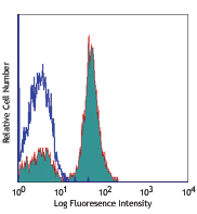
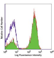
Human acute myeloid leukemia cell line KG1a was fixed, permeabilized, and stained with CD107b (clone H4B4) PE (filled histogram) or mouse IgG1, κ PE isotype control (open histogram). -

Human paraffin-embedded prostate tissue slice was stained with purified anti-human CD107b (clone H4B4) followed by goat anti-mouse IgG (clone Poly4053) DyLight™ 488 (green) and anti-human CD44 (clone IM7) Alexa Fluor® 594 (red). The nuclei were counterstained with DAPI (blue). The image was captured with a 10X objective. See additional supplemental data for detailed information. -

ICC staining of purified anti-LAMP-2 antibody (clone H4B4) on Hela cells. The cells were fixed with 4% PFA, permeabilized with 0.1% Triton X-100, and blocked with 2% normal goat serum and 0.02% BSA. The cells were then stained with 5 µg/ml of the primary antibody overnight at 4°C, followed by incubation with Alexa Fluor® 594 goat anti-Mouse IgG (Cat. No. 405326) for one hour at room temperature. Cells were counterstained with Phalloidin™ Green 488 (Cat. No. 424201) and DAPI to visualize actin filaments and nuclei, respectively. The images were captured with a 60X objective. Scale bar: 50 µm. -

Western blot of purified anti-LAMP-2 antibody (clone H4B4). Lane 1: Molecular weight marker; Lane 2: 20 µg of Hela cell lysate; Lane 3: 20 µg of NIH3T3 cell lysate; Lane 4: 20 µg of human brain lysates; Lane 5: 20 µg of mouse brain lysates; Lane 6: 10 ng human recombinant LAMP-2. The blot was incubated with 1 µg/ml of the primary antibody overnight at 4°C, followed by incubation with HRP-labeled goat anti-mouse IgG (Cat. No. 405306). Anti-β-actin antibody was used as the loading control (Cat. No. 643807). Enhanced chemiluminescence was used as the detection system.
| Cat # | Size | Price | Quantity Check Availability | Save | ||
|---|---|---|---|---|---|---|
| 354301 | 25 µg | 59 CHF | ||||
| 354302 | 100 µg | 114 CHF | ||||
CD107b, also known as LAMP-2, is a 105 kD, highly gylcosylated, type I transmembrane protein. CD107b is expressed in lysosomal/endosomal membranes in nearly all cells, and on the surface of activated platelets, activated lymphocytes and some tumor cell lines. LAMP-2 is known to have roles in cell adhesion and cellular homeostasis, including autophagocytosis and antigen presentation.
Product DetailsProduct Details
- Verified Reactivity
- Human
- Antibody Type
- Monoclonal
- Host Species
- Mouse
- Immunogen
- Adult human adherent spleen cells
- Formulation
- Phosphate-buffered solution, pH 7.2, containing 0.09% sodium azide.
- Preparation
- The antibody was purified by affinity chromatography.
- Concentration
- 0.5 mg/ml
- Storage & Handling
- The antibody solution should be stored undiluted between 2°C and 8°C.
- Application
-
ICFC - Quality tested
IHC-P, ICC, WB - Verified - Recommended Usage
-
Each lot of this antibody is quality control tested by intracellular immunofluorescent staining with flow cytometric analysis. For flow cytometric staining, the suggested use of this reagent is ≤ 0.5 µg per million cells in 100 µl volume. For immunohistochemical staining on formalin-fixed paraffin-embedded tissue sections, the suggested use of this reagent is 5.0 - 10 µg per ml. For Western blotting, the suggested use of this reagent is 1.0 - 5.0 µg per ml. For immunocytochemistry, a concentration range of 5.0 - 10 μg per ml is recommended. It is recommended that the reagent be titrated for optimal performance for each application.
- Application Notes
-
Additional reported applications (for the relevant formats) include: immunohistochemical staining of frozen glomeruli2 and immunofluorescent staining of neutrophils2,3.
This antibody is specific to human LAMP-2. Positive control: Hela cells; LAMP-2 molecular weight appears to be at ~110 kDa on the gel due to high glycosylation. -
Application References
(PubMed link indicates BioLegend citation) -
- Chen J, et al. 1985. J. Biol. Chem. 101:85.
- Kain R, et al. 2008. Nat. Med. 14:1088. (IF, IHC)
- Roark EA, et al. 2008. PLoS ONE 3:e3538. (IF)
- Product Citations
-
- RRID
-
AB_11204081 (BioLegend Cat. No. 354301)
AB_11204245 (BioLegend Cat. No. 354302)
Antigen Details
- Structure
- LAMP-2 is a 410 amino acid protein with a molecular mass of 45 kD.
- Distribution
-
Lysosomal/endosomal membranes in nearly all cells; surface of activated platelets, activated lymphocytes and some tumor cell lines
- Function
- Adhesion, cellular homeostatis, including autophagocytosis, antigen presentation
- Cell Type
- B cells, Lymphocytes, Platelets
- Biology Area
- Cell Adhesion, Cell Biology, Cell Motility/Cytoskeleton/Structure, Immunology, Innate Immunity, Neurodegeneration, Neuroscience, Protein Trafficking and Clearance
- Molecular Family
- Adhesion Molecules, CD Molecules
- Antigen References
-
1. Chen J, et al. 1985. J. Biol. Chem. 101:85.
2. Kain R, et al. 2008. Nat. Med. 14:1088.
3. Roark EA, et al. 2008. PLoS ONE 3:e3538. - Gene ID
- 3920 View all products for this Gene ID
- UniProt
- View information about CD107b on UniProt.org


Related FAQs
Other Formats
View All CD107b Reagents Request Custom Conjugation| Description | Clone | Applications |
|---|---|---|
| FITC anti-human CD107b (LAMP-2) | H4B4 | ICFC |
| PE anti-human CD107b (LAMP-2) | H4B4 | ICFC |
| Purified anti-human CD107b (LAMP-2) | H4B4 | ICFC,IHC-P,ICC,WB |
| Alexa Fluor® 594 anti-human CD107b (LAMP-2) | H4B4 | IHC-P |
| Alexa Fluor® 647 anti-human CD107b (LAMP-2) | H4B4 | ICFC,IHC-P |
| Brilliant Violet 421™ anti-human CD107b (LAMP-2) | H4B4 | ICFC,IHC-P |
Customers Also Purchased
Compare Data Across All Formats
This data display is provided for general comparisons between formats.
Your actual data may vary due to variations in samples, target cells, instruments and their settings, staining conditions, and other factors.
If you need assistance with selecting the best format contact our expert technical support team.
-
FITC anti-human CD107b (LAMP-2)

Human acute myeloid leukemia cell line KG1a was fixed, perme... -
PE anti-human CD107b (LAMP-2)

Human acute myeloid leukemia cell line KG1a was fixed, perme... -
Purified anti-human CD107b (LAMP-2)

Human acute myeloid leukemia cell line KG1a was fixed, perme... 
Human paraffin-embedded prostate tissue slice was stained wi... 
ICC staining of purified anti-LAMP-2 antibody (clone H4B4) o... 
Western blot of purified anti-LAMP-2 antibody (clone H4B4). ... -
Alexa Fluor® 594 anti-human CD107b (LAMP-2)

Human paraffin-embedded prostate tissue slices were prepared... -
Alexa Fluor® 647 anti-human CD107b (LAMP-2)

Human acute myeloid leukemia cell line KG1a was fixed, perme... 
Human paraffin-embedded prostate tissue slice were prepared ... -
Brilliant Violet 421™ anti-human CD107b (LAMP-2)

Human paraffin‐embedded prostate tissue slices were prepared... 
Human acute myeloid leukemia cell line KG1a was fixed, perme...


Follow Us