 Login / Register


- Clone
- H4A3 (See other available formats)
- Regulatory Status
- RUO
- Workshop
- P PR-63; BP 473; P P008
- Other Names
- Lysosome-Associated Membrane Protein 1, LGP-120, LAMP-1
- Isotype
- Mouse IgG1, κ
- Ave. Rating
- Submit a Review
- Product Citations
- publications

-

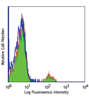
Thrombin-activated human peripheral blood platelets were stained with purified CD107a (clone H4A3) (red histogram) or purified mouse IgG1, κ (green histogram), followed by anti-mouse IgG FITC. -

ICC staining of purified anti-LAMP-1 antibody (clone H4A3) on Hela cells. The cells were fixed with 4% PFA, permeabilized with 0.1% Triton X-100, and blocked with 2% normal goat serum and 0.02% BSA. The cells were then stained with 5 µg/ml of the primary antibody overnight at 4°C, followed by incubation with Alexa Fluor® 594 goat anti-Mouse IgG (Cat. No. 405326) for one hour at room temperature. Cells were counterstained with Phalloidin™ Green 488 (Cat. No. 424201) and DAPI to visualize actin filaments and nuclei, respectively. The images were captured with a 60X objective. Scale bar: 50 µm. -

Western blot of purified anti-LAMP-1 antibody (clone H4A3). Lane 1: Molecular weight marker; Lane 2: 20 µg of Hela cell lysate; Lane 3: 20 µg of NIH3T3 cell lysate; Lane 4: 20 µg of human brain lysate; Lane 5: 20 µg of mouse brain lysate; Lane 6: 10 ng of human recombinant LAMP-1. The blot was incubated with 1 µg/ml of the primary antibody overnight at 4°C, followed by incubation with HRP-labeled goat anti-mouse IgG (Cat. No. 405306). Anti-β-actin antibody was used as the loading control (Cat. No. 643807). Enhanced chemiluminescence was used as the detection system. -

Formalin-fixed paraffin-embedded human tonsil treated with a citrate buffer pH6 and heat for antigen retrieval was stained with purified CD107a clone H4A3, conjugated and detected with a Cy5 conjugated CODEX™ oligonucleotide duplex (green). Data generated at Akoya Biosciences, Inc. using the CODEX™ technology. -

SeqIF™ (sequential immunofluorescence) staining on COMET™ of anti-CD107a (LAMP-1) (clone H4A3, yellow) on formalin-fixed paraffin-embedded human pancreatic carcinoma tissue at 5 µg/mL. Alexa Fluor™ Plus 647 Goat anti-Mouse IgG antibody (Lunaphore, Cat. No. DR647MS) was used as a secondary antibody. Nuclei were counterstained with DAPI (blue). Tissue underwent an all-in-one dewaxing and antigen retrieval preprocessing.
| Cat # | Size | Price | Quantity Check Availability | Save | ||
|---|---|---|---|---|---|---|
| 328601 | 25 µg | 59 CHF | ||||
| 328602 | 100 µg | 114 CHF | ||||
CD107a, also known as Lysosome-Associated Membrane Protein 1 (LAMP-1) or LGP-120, is a 110-140 kD type I membrane glycoprotein. Mature CD107a is heavily glycosylated from a 40 kD core protein. This molecule is located on the luminal side of lysosomes. Upon activation, CD107a is transferred to the cell membrane surface of activated platelets, activated lymphocytes, macrophages, epithelial cells, endothelial cells, and some tumor cells. CD107a has been suggested to play a role in the protection of lysosomal membrane from lysosomal hydrolases which is involved in cell adhesion and regulation of tumor metastasis, and mediates autoimmune disease progression. CD107a is a ligand for galaptin and E-selectin. Surface expression of LAMP-1 has been shown to correlate with CD8+ T cell and NK cell cytotoxicity.
Product DetailsProduct Details
- Verified Reactivity
- Human
- Reported Reactivity
- African Green, Baboon, Chimpanzee, Cynomolgus, Pigtailed Macaque, Rhesus
- Antibody Type
- Monoclonal
- Host Species
- Mouse
- Immunogen
- Human adult adherent peripheral blood cells
- Formulation
- Phosphate-buffered solution, pH 7.2, containing 0.09% sodium azide.
- Preparation
- The antibody was purified by affinity chromatography.
- Concentration
- 0.5 mg/ml
- Storage & Handling
- The antibody solution should be stored undiluted between 2°C and 8°C.
- Application
-
FC - Quality tested
ICC, WB, IHC-P - Verified
IP - Reported in the literature, not verified in house
SB - Community verified - Recommended Usage
-
Each lot of this antibody is quality control tested by immunofluorescent staining with flow cytometric analysis. For flow cytometric staining, the suggested use of this reagent is ≤ 2.0 µg per million cells in 100 µl volume. For Western blotting, the suggested use of this reagent is 1.0 - 5.0 µg per ml. For immunocytochemistry on formalin-fixed paraffin-embedded tissue sections, a concentration range of 5.0 - 10 μg per ml is recommended. It is recommended that the reagent be titrated for optimal performance for each application.
- Application Notes
-
Additional reported applications (for the relevant formats) include: Western blotting8, immunohistochemical staining2, immunofluorescence5,7, and immunoprecipitation5.
This antibody is specific to human LAMP-1. Positive control: Hela cells; LAMP-1 molecular weight appears to be at ~110 kDa on the gel due to high glycosylation. - Additional Product Notes
-
For the use of this antibody in spatial biology applications, we have partnered with Lunaphore Technologies for demonstration of our antibodies on the COMET™. The COMET™ platform is an automated, end-to-end spatial biology solution developed for rapid and flexible multiplex tissue profiling. More information on the COMET™ and a complete list of our antibodies that have been demonstrated on the COMET™ can be found here.
-
Application References
(PubMed link indicates BioLegend citation) -
- Misse D, et al. 1999. Blood 93:2454.
- Furuta K, et al. 2001. Am. J. Pathol. 159:449. (IHC)
- Watanabe A, et al. 2011. J. Biol. Chem. 286:10702. PubMed
- Baron Gaillard CL, et al. 2011. Mol. Cell. Biol. 22:5459. PubMed
- Hauck CR and Meyer TF. 1997. FEBS Lett. 405:86. (IF, IP)
- De Keersmaecker B, et al. 2012. J. Virol. 86:9351. PubMed
- Knodler LA, et al. 2010. P. Natl. Acad. Sci. USA. 107:17733. (IF)
- Oh J, et al. 2000. Hum. Mol. Genet. 9:375. (WB)
- Salio M, et al. 2013 PNAS. 110:4753. PubMed
- Product Citations
-
- RRID
-
AB_1134260 (BioLegend Cat. No. 328601)
AB_1134259 (BioLegend Cat. No. 328602)
Antigen Details
- Structure
- LAMP-1 is a 417 amino acid protein with a molecular mass of 45 kD.
- Distribution
-
Macrophages, epithelial cells, endothelial cells, some tumor cells; located on the luminal side of lysosomes or on the surface of cell membranes
- Function
- Protect lysosomal membrane from lysosomal hydrolases, adhesion
- Ligand/Receptor
- Galaptin
- Cell Type
- Endothelial cells, Epithelial cells, Macrophages
- Biology Area
- Cell Biology, Immunology, Neurodegeneration, Neuroscience, Protein Trafficking and Clearance
- Molecular Family
- Adhesion Molecules, CD Molecules
- Antigen References
-
1. Sarafian V, et al. 2006. Arch. Dermatol. Res. 298:7381.
2. Schlossman SF, et al. 1995. Leukocyte Typing V:White Cell Differentiation Antigens. New York:Oxford University Press.
3. Sawada R, et al. 1993. J. Biol. Chem. 268:12675.
4. Chen JW, et al. 1988. J. Biol. Chem. 263:8754.
5. Chen JW, et al. 1986. Biochem. Soc. Symp. 51:97112. - Gene ID
- 3916 View all products for this Gene ID
- UniProt
- View information about CD107a on UniProt.org


Related FAQs
- If an antibody clone has been previously successfully used in IBEX in one fluorescent format, will other antibody formats work as well?
-
It’s likely that other fluorophore conjugates to the same antibody clone will also be compatible with IBEX using the same sample fixation procedure. Ultimately a directly conjugated antibody’s utility in fluorescent imaging and IBEX may be specific to the sample and microscope being used in the experiment. Some antibody clone conjugates may perform better than others due to performance differences in non-specific binding, fluorophore brightness, and other biochemical properties unique to that conjugate.
- Will antibodies my lab is already using for fluorescent or chromogenic IHC work in IBEX?
-
Fundamentally, IBEX as a technique that works much in the same way as single antibody panels or single marker IF/IHC. If you’re already successfully using an antibody clone on a sample of interest, it is likely that clone will have utility in IBEX. It is expected some optimization and testing of different antibody fluorophore conjugates will be required to find a suitable format; however, legacy microscopy techniques like chromogenic IHC on fixed or frozen tissue is an excellent place to start looking for useful antibodies.
- Are other fluorophores compatible with IBEX?
-
Over 18 fluorescent formats have been screened for use in IBEX, however, it is likely that other fluorophores are able to be rapidly bleached in IBEX. If a fluorophore format is already suitable for your imaging platform it can be tested for compatibility in IBEX.
- The same antibody works in one tissue type but not another. What is happening?
-
Differences in tissue properties may impact both the ability of an antibody to bind its target specifically and impact the ability of a specific fluorophore conjugate to overcome the background fluorescent signal in a given tissue. Secondary stains, as well as testing multiple fluorescent conjugates of the same clone, may help to troubleshoot challenging targets or tissues. Using a reference control tissue may also give confidence in the specificity of your staining.
- How can I be sure the staining I’m seeing in my tissue is real?
-
In general, best practices for validating an antibody in traditional chromogenic or fluorescent IHC are applicable to IBEX. Please reference the Nature Methods review on antibody based multiplexed imaging for resources on validating antibodies for IBEX.
Other Formats
View All CD107a Reagents Request Custom ConjugationCustomers Also Purchased
Compare Data Across All Formats
This data display is provided for general comparisons between formats.
Your actual data may vary due to variations in samples, target cells, instruments and their settings, staining conditions, and other factors.
If you need assistance with selecting the best format contact our expert technical support team.
-
Biotin anti-human CD107a (LAMP-1)

Thrombin-activated human peripheral blood platelets were sta... -
Purified anti-human CD107a (LAMP-1)

Thrombin-activated human peripheral blood platelets were sta... 
ICC staining of purified anti-LAMP-1 antibody (clone H4A3) o... 
Western blot of purified anti-LAMP-1 antibody (clone H4A3). ... 
Formalin-fixed paraffin-embedded human tonsil treated with a... 
SeqIF™ (sequential immunofluorescence) staining on COMET™ of... -
FITC anti-human CD107a (LAMP-1)

Thrombin-activated human peripheral blood platelets were sta... -
PE anti-human CD107a (LAMP-1)

Thrombin-activated human peripheral blood platelets were sta... -
Alexa Fluor® 488 anti-human CD107a (LAMP-1)

Thrombin-activated human peripheral blood platelets were sta... -
Alexa Fluor® 647 anti-human CD107a (LAMP-1)

Thrombin-activated human peripheral blood platelets were sta... -
PerCP/Cyanine5.5 anti-human CD107a (LAMP-1)

PMA+Ionomycin stimulated (4 hours) human peripheral blood mo... -
APC anti-human CD107a (LAMP-1)

Thrombin-activated human peripheral blood platelets were sta... -
Pacific Blue™ anti-human CD107a (LAMP-1)

Thrombin-activated human peripheral blood platelets were sta... -
Brilliant Violet 421™ anti-human CD107a (LAMP-1)

Thrombin-activated human peripheral blood platelets were sta... -
PE/Cyanine7 anti-human CD107a (LAMP-1)

Thrombin-activated human peripheral blood platelets were sta... -
APC/Cyanine7 anti-human CD107a (LAMP-1)

PMA+Ionomycin stimulated (6 hours) human peripheral blood mo... -
Brilliant Violet 510™ anti-human CD107a (LAMP-1)

Thrombin-activated human peripheral blood platelets were sta... -
Brilliant Violet 605™ anti-human CD107a (LAMP-1)

Thrombin-activated human peripheral blood platelets were sta... -
Purified anti-human CD107a (LAMP-1) (Maxpar® Ready)

Human PBMCs were incubated for 5 hours in media alone (top) ... 
-
Brilliant Violet 650™ anti-human CD107a (LAMP-1)

Thrombin-activated human peripheral blood platelets were sta... -
Brilliant Violet 711™ anti-human CD107a (LAMP-1)

Thrombin-activated human peripheral blood platelets were sta... -
PerCP anti-human CD107a (LAMP-1)

Thrombin-activated human peripheral blood platelets were sta... -
Brilliant Violet 785™ anti-human CD107a (LAMP-1)

Thrombin-activated human peripheral blood platelets were sta... -
PE/Dazzle™ 594 anti-human CD107a (LAMP-1)

Thrombin-activated human peripheral blood platelets were sta... -
TotalSeq™-A0155 anti-human CD107a (LAMP-1)
-
TotalSeq™-C0155 anti-human CD107a (LAMP-1)
-
TotalSeq™-B0155 anti-human CD107a (LAMP-1)
-
APC/Fire™ 750 anti-human CD107a (LAMP-1) Antibody

Thrombin-activated human peripheral blood platelets were sta... -
PE/Cyanine5 anti-human CD107a (LAMP-1)

Thrombin-activated human peripheral blood platelets were sta...


Follow Us