 Login / Register


- Clone
- Poly4064 (See other available formats)
- Regulatory Status
- RUO
- Isotype
- Donkey Polyclonal Ig
- Ave. Rating
- Submit a Review
- Product Citations
- publications

-

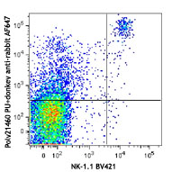
C57BL/6 splenocytes were stained with NK-1.1 APC and purified rabbit anti-Asialo-GM1, followed by donkey anti-rabbit IgG (clone Poly4064) PE (top image).
| Cat # | Size | Price | Quantity Check Availability | Save | ||
|---|---|---|---|---|---|---|
| 406421 | 100 µg | 104 CHF | ||||
This polyclonal donkey anti-rabbit IgG antibody reacts with the heavy chains of rabbit IgG and with the light chains common to most rabbit immunoglobulins. No cross-reactivity has been detected against non-immunoglobulin serum proteins. This antibody has been solid-phase absorbed to ensure minimal cross-reaction with rat, human, bovine, horse, and mouse immunoglobulins, but it may cross-react with other subclasses of rabbit immunoglobulins.
Product DetailsProduct Details
- Verified Reactivity
- Rabbit
- Antibody Type
- Polyclonal
- Host Species
- Donkey
- Formulation
- Phosphate-buffered solution, pH 7.2, containing 0.09% sodium azide.
- Preparation
- The antibody was purified by affinity chromatography and conjugated with PE under optimal conditions.
- Concentration
- 0.2 mg/ml
- Storage & Handling
- The antibody solution should be stored undiluted between 2°C and 8°C, and protected from prolonged exposure to light. Do not freeze.
- Application
-
FC - Quality tested
- Recommended Usage
-
Each lot of this antibody is quality control tested by immunofluorescent staining with flow cytometric analysis. For flow cytometric staining, the suggested use of this reagent is ≤0.125 µg per million cells in 100 µl volume. It is recommended that the reagent be titrated for optimal performance for each application.
- Excitation Laser
-
Blue Laser (488 nm)
Green Laser (532 nm)/Yellow-Green Laser (561 nm)
- Application Notes
-
This conjugated polyclonal donkey anti-rabbit IgG antibody is useful for immunofluorescent staining for flow cytometry, or microscopy.
- Product Citations
-
- RRID
-
AB_2563484 (BioLegend Cat. No. 406421)


Related FAQs
- What type of PE do you use in your conjugates?
- We use R-PE in our conjugates.
Other Formats
View All Rabbit IgG Reagents Request Custom Conjugation| Description | Clone | Applications |
|---|---|---|
| HRP Donkey anti-rabbit IgG (minimal x-reactivity) | Poly4064 | ELISA,IHC,WB |
| Cyanine3 Donkey anti-rabbit IgG (minimal x-reactivity) | Poly4064 | ICC |
| FITC Donkey anti-rabbit IgG (minimal x-reactivity) | Poly4064 | ICC,FC |
| Brilliant Violet 421™ Donkey anti-rabbit IgG (minimal x-reactivity) | Poly4064 | ICC,FC |
| DyLight™ 488 Donkey anti-rabbit IgG (minimal x-reactivity) | Poly4064 | ICC,FC |
| DyLight™ 649 Donkey anti-rabbit IgG (minimal x-reactivity) | Poly4064 | ICC,FC |
| Alexa Fluor® 647 Donkey anti-rabbit IgG (minimal x-reactivity) | Poly4064 | FC |
| Alexa Fluor® 488 Donkey anti-rabbit IgG (minimal x-reactivity) | Poly4064 | FC |
| PE Donkey anti-rabbit IgG (minimal x-reactivity) | Poly4064 | FC |
| Brilliant Violet 510™ Donkey anti-rabbit IgG (minimal x-reactivity) | Poly4064 | FC |
| Alexa Fluor® 594 Donkey anti-rabbit IgG (minimal x-reactivity) | Poly4064 | ICC |
| Alexa Fluor® 555 Donkey anti-rabbit IgG (minimal x-reactivity) | Poly4064 | ICC |
Customers Also Purchased
Compare Data Across All Formats
This data display is provided for general comparisons between formats.
Your actual data may vary due to variations in samples, target cells, instruments and their settings, staining conditions, and other factors.
If you need assistance with selecting the best format contact our expert technical support team.
-
HRP Donkey anti-rabbit IgG (minimal x-reactivity)
-
Cyanine3 Donkey anti-rabbit IgG (minimal x-reactivity)

HeLa cells were fixed with ice cold methanol for six minutes... -
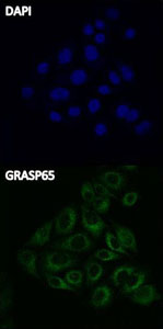
FITC Donkey anti-rabbit IgG (minimal x-reactivity)

Hela cells were stained with anti-GRASP65 (Poly6210) and sec... -
Brilliant Violet 421™ Donkey anti-rabbit IgG (minimal x-reactivity)

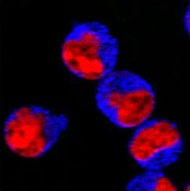
293E cells were stained by purified anti-Tubulin-gamma (clon... -
DyLight™ 488 Donkey anti-rabbit IgG (minimal x-reactivity)

293T cells were stained with anti-Tubulin-γ and secondarily ... -
DyLight™ 649 Donkey anti-rabbit IgG (minimal x-reactivity)
-
Alexa Fluor® 647 Donkey anti-rabbit IgG (minimal x-reactivity)

C57/BL6 splenocytes were stained with NK-1.1 Brilliant Viole... -
Alexa Fluor® 488 Donkey anti-rabbit IgG (minimal x-reactivity)

C57BL/6 splenocytes were stained with NK-1.1 Brilliant Viole... -
PE Donkey anti-rabbit IgG (minimal x-reactivity)

C57BL/6 splenocytes were stained with NK-1.1 APC and purifie... -
Brilliant Violet 510™ Donkey anti-rabbit IgG (minimal x-reactivity)

C57BL/6 splenocytes were stained with NK-1.1 APC and purifie... -
Alexa Fluor® 594 Donkey anti-rabbit IgG (minimal x-reactivity)

HeLa cells were fixed with 2% paraformaldehyde (PFA) for 10 ... -
Alexa Fluor® 555 Donkey anti-rabbit IgG (minimal x-reactivity)

HeLa cells were fixed with 1% paraformaldehyde for ten minut...


Follow Us