 Login / Register


- Clone
- M-T271 (See other available formats)
- Regulatory Status
- RUO
- Workshop
- V 5T CD27.03
- Other Names
- S152, T14, TNFRSF7
- Isotype
- Mouse IgG1, κ
- Ave. Rating
- Submit a Review
- Product Citations
- publications

-

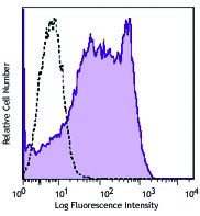
Human peripheral blood lymphocytes were stained with anti-human CD27 (clone M-T271) PE/Cyanine5 (filled histogram) or mouse IgG1, κ PE/Cyanine5 isotype control (open histogram).
| Cat # | Size | Price | Quantity Check Availability | Save | ||
|---|---|---|---|---|---|---|
| 356437 | 25 tests | 183 CHF | ||||
| 356438 | 100 tests | 370 CHF | ||||
CD27 is a 50-55 kD type I membrane protein also known as S152 and T14. It is a lymphocyte-specific member of the TNF-receptor superfamily. CD27 is expressed on medullary thymocytes, virtually all mature T cells, some B cells, and NK cells. CD27 binds to CD70, and plays a role in costimulation of T cell activation and regulation of B cell differentiation and proliferation. The cytoplasmic domains of CD27 have also been shown to interact with TRAF2 and TRAF5 to elicit NF-κB and SAPK/JNK activation.
Product DetailsProduct Details
- Verified Reactivity
- Human
- Reported Reactivity
- Baboon, Cynomolgus, Rhesus
- Antibody Type
- Monoclonal
- Host Species
- Mouse
- Immunogen
- Human T cells from a T-ALL patient.
- Formulation
- Phosphate-buffered solution, pH 7.2, containing 0.09% sodium azide and BSA (origin USA)
- Preparation
- The antibody was purified by affinity chromatography and conjugated with PE/Cyanine5 under optimal conditions.
- Concentration
- Lot-specific (to obtain lot-specific concentration and expiration, please enter the lot number in our Certificate of Analysis online tool.)
- Storage & Handling
- The antibody solution should be stored undiluted between 2°C and 8°C, and protected from prolonged exposure to light. Do not freeze.
- Application
-
FC - Quality tested
- Recommended Usage
-
Each lot of this antibody is quality control tested by immunofluorescent staining with flow cytometric analysis. For flow cytometric staining, the suggested use of this reagent is 5 µL per million cells in 100 µL staining volume or 5 µL per 100 µL of whole blood. It is recommended that the reagent be titrated for optimal performance for each application.
- Excitation Laser
-
Blue Laser (488 nm)
Green Laser (532 nm)/Yellow-Green Laser (561 nm)
- Application Notes
-
Additional reported applications (for the relevant formats) include: immunohistochemical staining of formalin-fixed paraffin-embedded frozen tissue sections1, immunofluorescent staining2, and ELISA3.
-
Application References
(PubMed link indicates BioLegend citation) -
- Ma S, et al. 2011. J. Virol. 85:165. (IHC)
- Manzo A, et al. 2008. Arthritis Rheum. 11:3377. (IF)
- Kato K, et al. 2007. Exp. Hematol. 35:434. (ELISA)
- RRID
-
AB_2894486 (BioLegend Cat. No. 356437)
AB_2894486 (BioLegend Cat. No. 356438)
Antigen Details
- Structure
- TNF-R superfamily, type I transmembrane glycoprotein, 50-55 kD
- Distribution
-
Medullary thymocytes, T and B cell subsets, NK cells
- Interaction
- CD27 binds to CD70
- Ligand/Receptor
- CD70
- Cell Type
- B cells, NK cells, T cells, Thymocytes
- Biology Area
- Costimulatory Molecules, Immunology
- Molecular Family
- CD Molecules
- Antigen References
-
1. Knapp W, et al. 1989. Leucocyte Typing IV: White Cell Differentiation Antigens. Oxford University Press.
2. Schlossman S, et al. 1995. Leucocyte Typing V: White Cell Differentiation Antigens. Oxford University Press.
3. Hintzen R, et al. 1994. Immunol. Today 15:307.
4. Agematsu K, et al. 1995. J. Immunol. 154:3627. - Gene ID
- 939 View all products for this Gene ID
- UniProt
- View information about CD27 on UniProt.org
Related Pages & Pathways
Pathways


Other Formats
View All CD27 Reagents Request Custom ConjugationCustomers Also Purchased
Compare Data Across All Formats
This data display is provided for general comparisons between formats.
Your actual data may vary due to variations in samples, target cells, instruments and their settings, staining conditions, and other factors.
If you need assistance with selecting the best format contact our expert technical support team.
-
Purified anti-human CD27

Human peripheral blood lymphocytes were stained with purifie... -
FITC anti-human CD27

Human peripheral blood lymphocytes were stained with CD27 (c... -
PE anti-human CD27

Human peripheral blood lymphocytes were stained with CD27 (c... 
Human peripheral blood was stained with CD27 (clone M-T271) ... -
PerCP/Cyanine5.5 anti-human CD27

Human peripheral blood lymphocytes were stained with CD27 (c... -
APC anti-human CD27

Human peripheral blood lymphocytes were stained with CD27 (c... -
PE/Cyanine7 anti-human CD27

Human peripheral blood lymphocytes were stained with CD27 (c... -
Pacific Blue™ anti-human CD27

Human peripheral blood lymphocytes were stained with CD27 (c... -
Alexa Fluor® 700 anti-human CD27

Human peripheral blood lymphocytes were stained with CD27 (c... -
Brilliant Violet 421™ anti-human CD27

Human peripheral blood lymphocytes were stained with CD27 (c... -
Brilliant Violet 510™ anti-human CD27

Human peripheral blood lymphocytes were stained with CD27 (c... -
PE/Dazzle™ 594 anti-human CD27

Human peripheral blood lymphocytes were stained with CD27 (c... -
APC/Cyanine7 anti-human CD27

Human peripheral blood lymphocytes were stained with CD27 (c... -
Biotin anti-human CD27

Human peripheral blood lymphocytes were stained with biotiny... -
APC/Fire™ 750 anti-human CD27

Human peripheral blood lymphocytes were stained with CD27 (c... -
Brilliant Violet 711™ anti-human CD27

Human peripheral blood lymphocytes were stained with CD27 (c... -
PerCP anti-human CD27

Human peripheral blood lymphocytes were stained with CD27 (c... -
Alexa Fluor® 647 anti-human CD27

Human peripheral blood lymphocytes were stained with CD27 (c... -
KIRAVIA Blue 520™ anti-human CD27

Human Peripheral blood lymphocytes were stained with CD3 APC... -
PE/Cyanine5 anti-human CD27

Human peripheral blood lymphocytes were stained with anti-hu... -
PE/Cyanine7 anti-human CD27

Typical results from human peripheral blood lymphocytes stai... -
APC anti-human CD27

Typical results from human peripheral blood lymphocytes stai... -
PerCP/Cyanine5.5 anti-human CD27

Typical results from human peripheral blood lymphocytes stai... -
PE anti-human CD27

Typical result from human peripheral blood lymphocytes stain... -
FITC anti-human CD27

Typical results from human peripheral blood lymphocytes stai... -
Brilliant Violet 650™ anti-human CD27

Human peripheral blood lymphocytes were stained with anti-hu... -
Pacific Blue™ anti-human CD27

Typical result from human peripheral lymphocytes stained eit... -
Spark Violet™ 423 anti-human CD27

Human peripheral blood lymphocytes were stained with anti-hu... -
GMP PE/Cyanine7 anti-human CD27

Typical results from human peripheral blood lymphocytes stai... -
Spark Red™ 718 anti-human CD27 (Flexi-Fluor™)


Follow Us