 Login / Register


- Clone
- 14G2a (See other available formats)
- Regulatory Status
- RUO
- Other Names
- Disialoganglioside GD2, Ganglioside G2
- Isotype
- Mouse IgG2a, κ
- Ave. Rating
- Submit a Review
- Product Citations
- publications

-

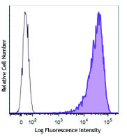
Human melanoma cell line M21 was stained with ganglioside GD2 (clone 14G2a) PE (filled histogram) or mouse IgG2a, κ PE isotype control (open histogram).
| Cat # | Size | Price | Quantity Check Availability | Save | ||
|---|---|---|---|---|---|---|
| 357303 | 25 tests | 153 CHF | ||||
| 357304 | 100 tests | 384 CHF | ||||
Ganglioside GD2 is a sialic acid-containing glycosphingolipid involved in cell attachment to the extracellular matrix. Expression of GD2 in normal tissue is restricted to cells from the central nervous system, peripheral nerves, skin melanocytes, and mesenchymal stem cells. However GD2 is highly expressed by tumors of neuro-ectodermal origin such as melanomas, gliomas, neuroblastomas, and small cell lung carcinoma. GD2 has been proposed as a marker for some cancer stem cells.
Product DetailsProduct Details
- Verified Reactivity
- Human
- Antibody Type
- Monoclonal
- Host Species
- Mouse
- Immunogen
- Neuroblastoma cell line LAN-1
- Formulation
- Phosphate-buffered solution, pH 7.2, containing 0.09% sodium azide and BSA (origin USA)
- Preparation
- The antibody was purified by affinity chromatography and conjugated with PE under optimal conditions.
- Concentration
- Lot-specific (to obtain lot-specific concentration and expiration, please enter the lot number in our Certificate of Analysis online tool.)
- Storage & Handling
- The antibody solution should be stored undiluted between 2°C and 8°C, and protected from prolonged exposure to light. Do not freeze.
- Application
-
FC - Quality tested
- Recommended Usage
-
Each lot of this antibody is quality control tested by immunofluorescent staining with flow cytometric analysis. For flow cytometric staining, the suggested use of this reagent is 5 µl per million cells in 100 µl staining volume or 5 µl per 100 µl of whole blood.
- Excitation Laser
-
Blue Laser (488 nm)
Green Laser (532 nm)/Yellow-Green Laser (561 nm)
- Application Notes
-
Clone 14G2a is an isotype switch variant from parental hybridoma 14.18 (IgG3)1. Additional reported applications (for the relevant formats) include: inducing apoptosis and enhancing cytotoxicity of chemotherapeutic drugs in the neuroblastoma cell line 2. The LEAF™ or Ultra-LEAF™ purified antibody is recommended for use in cytotoxic and apoptotic studies (contact our custom solutions team). This clone has also been published as 14.G2a.
-
Application References
(PubMed link indicates BioLegend citation) -
- Mujoo K, et al. 1989. Cancer Res. 49:2857. (Cyt)
- Kowalczyk A, et al. 2009. Cancer Lett. 281:171. (Apop, Cyt)
- Battula VL, et al. 2012. J. Clin. Invest. 122:2066. (FC)
- Product Citations
-
- RRID
-
AB_2561884 (BioLegend Cat. No. 357303)
AB_2561885 (BioLegend Cat. No. 357304)
Antigen Details
- Distribution
-
Neurons, mesenchymal stem cells, highly expressed in tumors of neuro-ectodermal origin, such as melanomas, gliomas, neuroblastomas, and small cell lung carcinoma
- Function
- Cell attachment to extracellular matrix
- Cell Type
- Mesenchymal cells, Neurons
- Biology Area
- Apoptosis/Tumor Suppressors/Cell Death, Cell Adhesion, Cell Biology, Immunology, Neuroscience
- Molecular Family
- Adhesion Molecules
- Antigen References
-
1. Tarek N, et al. 2012. J. Clin. Invest. 122:3260.
2. Matthay KK, et al. 2012. Clin. Cancer Res. 18:2740.
3. Navid F, et al. 2010. Curr. Cancer Drug Targets. 10:200. - Gene ID
- 2583 View all products for this Gene ID
- UniProt
- View information about Ganglioside GD2 on UniProt.org


Related FAQs
- What type of PE do you use in your conjugates?
- We use R-PE in our conjugates.
Other Formats
View All Ganglioside GD2 Reagents Request Custom Conjugation| Description | Clone | Applications |
|---|---|---|
| Purified anti-human Ganglioside GD2 | 14G2a | FC |
| PE anti-human Ganglioside GD2 | 14G2a | FC |
| APC anti-human Ganglioside GD2 | 14G2a | FC |
| PE/Cyanine7 anti-human Ganglioside GD2 | 14G2a | FC |
| Biotin anti-human Ganglioside GD2 | 14G2a | FC |
| PerCP/Cyanine5.5 anti-human Ganglioside GD2 | 14G2a | FC |
| FITC anti-human Ganglioside GD2 | 14G2a | FC |
| Brilliant Violet 510™ anti-human Ganglioside GD2 | 14G2a | FC |
| Alexa Fluor® 647 anti-human Ganglioside GD2 | 14G2a | FC |
| PE/Dazzle™ 594 anti-human Ganglioside GD2 | 14G2a | FC |
| APC/Fire™ 750 anti-human Ganglioside GD2 | 14G2a | FC |
| APC/Cyanine7 anti-human Ganglioside GD2 | 14G2a | FC |
| Brilliant Violet 421™ anti-human Ganglioside GD2 | 14G2a | FC |
| TotalSeq™-C1456 anti-human Ganglioside GD2 | 14G2a | PG |
| TotalSeq™-A1456 anti-human Ganglioside GD2 | 14G2a | PG |
Customers Also Purchased
Compare Data Across All Formats
This data display is provided for general comparisons between formats.
Your actual data may vary due to variations in samples, target cells, instruments and their settings, staining conditions, and other factors.
If you need assistance with selecting the best format contact our expert technical support team.
-
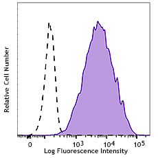
Purified anti-human Ganglioside GD2

Human melanoma cell line M21 was stained with purified gangl... -
PE anti-human Ganglioside GD2

Human melanoma cell line M21 was stained with ganglioside GD... -
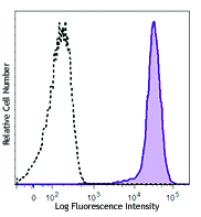
APC anti-human Ganglioside GD2

Human melanoma cell line M21 was stained with ganglioside GD... -
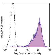
PE/Cyanine7 anti-human Ganglioside GD2

Human melanoma cell line M21 was stained with ganglioside GD... -
Biotin anti-human Ganglioside GD2

Human melanoma cell line M21 was stained with Ganglioside GD... -
PerCP/Cyanine5.5 anti-human Ganglioside GD2

Human melanoma cell line M21 was stained with Ganglioside GD... -
FITC anti-human Ganglioside GD2

Human melanoma cell line M21 was stained with Ganglioside GD... -
Brilliant Violet 510™ anti-human Ganglioside GD2

Human melanoma cell line M21 was stained with Ganglioside GD... -
Alexa Fluor® 647 anti-human Ganglioside GD2

Human melanoma cell line M21 was stained with Ganglioside GD... -
PE/Dazzle™ 594 anti-human Ganglioside GD2

Human melanoma cell line M21 was stained with Ganglioside GD... -
APC/Fire™ 750 anti-human Ganglioside GD2

Human melanoma cell line M21 was stained with Ganglioside GD... -
APC/Cyanine7 anti-human Ganglioside GD2

Human melanoma cell line M21 was stained with Ganglioside GD... -
Brilliant Violet 421™ anti-human Ganglioside GD2

Human melanoma cell line M21 was stained with anti-human Gan... -
TotalSeq™-C1456 anti-human Ganglioside GD2
-
TotalSeq™-A1456 anti-human Ganglioside GD2


Follow Us