 Login / Register


- Clone
- HI29 (See other available formats)
- Regulatory Status
- RUO
- Workshop
- IV N18
- Other Names
- OX44, MOX44
- Isotype
- Mouse IgG1, κ
- Ave. Rating
- Submit a Review
- Product Citations
- publications

-

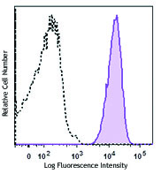
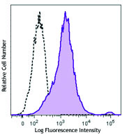
Human peripheral blood lymphocytes stained with HI29 PE
| Cat # | Size | Price | Quantity Check Availability | Save | ||
|---|---|---|---|---|---|---|
| 325406 | 100 tests | 281 CHF | ||||
CD53 is a 35-42 kD type III tetraspan membrane protein. It is expressed on all leukocytes, but not on platelets or erythrocytes. CD53 is thought to be involved in signal transduction. It has been shown to interact with a number of proteins including IL-4, CD20, CD2, CD9, IL-2, VLA-4, and CD4. Cross-linking of CD53 promotes B cell activation. The CD53 antigen is highly glycosylated and treatment with endoglycosidase F reduces the apparent molecular weight of this protein by approximately 25 kD. The HI29 antibody has been shown to be useful for flow cytometry, and immunohistochemistry (frozen).
Product DetailsProduct Details
- Verified Reactivity
- Human
- Reported Reactivity
- Chimpanzee
- Antibody Type
- Monoclonal
- Host Species
- Mouse
- Formulation
- Phosphate-buffered solution, pH 7.2, containing 0.09% sodium azide and BSA (origin USA)
- Preparation
- The antibody was purified by affinity chromatography, and conjugated with PE under optimal conditions.
- Concentration
- Lot-specific (to obtain lot-specific concentration and expiration, please enter the lot number in our Certificate of Analysis online tool.)
- Storage & Handling
- The antibody solution should be stored undiluted between 2°C and 8°C, and protected from prolonged exposure to light. Do not freeze.
- Application
-
FC - Quality tested
- Recommended Usage
-
Each lot of this antibody is quality control tested by immunofluorescent staining with flow cytometric analysis. For flow cytometric staining, the suggested use of this reagent is 5 µl per million cells in 100 µl staining volume or 5 µl per 100 µl of whole blood.
- Excitation Laser
-
Blue Laser (488 nm)
Green Laser (532 nm)/Yellow-Green Laser (561 nm)
- Application Notes
-
Additional reported applications (for the relevant formats) include: immunohistochemistry of acetone-fixed frozen tissue sections.
-
Application References
(PubMed link indicates BioLegend citation) -
- Mollinedo F, et al. 1997. Clin. and Diag. lab. Immunol. 4:229.
- Cao L, et al. 1997. Immunobiology 197(1):70.
- Amiot M. 1990. J. Immunol. 145:4322.
- Vanherberghen B, et al. 2004. Proc Natl Acad Sci USA. 101(48):16873.
- Yoshino N, et al. 2000. Exp. Anim. (Tokyo) 49:97. (FC)
- Product Citations
-
- RRID
-
AB_2075725 (BioLegend Cat. No. 325406)
Antigen Details
- Structure
- Cell surface protein, tetraspan family, four membrane spanning hydrophobic domains, 35-42 kD
- Distribution
-
Hematopoietic cells including neutrophils, monocytes, B cells, T cells (single positive thymocytes and peripheral T cells), and eosinophils. Not expressed on platelets, erythrocytes, and non-hematopoietic cells
- Function
- Signal transduction, cross-linking on B cells causes activation
- Interaction
- IL-4, CD20, CD2, CD9, IL-2, integrin α4, integrin β1, CD4
- Modification
- Heavily glycosylated, treatment with endoglycosidase F reduces apparent molecular weight by 25 kD
- Cell Type
- B cells, Eosinophils, Neutrophils
- Biology Area
- Costimulatory Molecules, Immunology
- Molecular Family
- CD Molecules
- Antigen References
-
1. Amiot M. 1990. J. Immunol. 145:4322.
2. Angelisova P, et al. 1990. Immunogenetics 32:281.
3. Olweus J, et al. 1993. J. Immunol. 153:4997.
4. Leukocyte Typing IV. Knapp W, et al. (Eds) Oxford University Press (1989) - Gene ID
- 963 View all products for this Gene ID
- UniProt
- View information about CD53 on UniProt.org
Related Pages & Pathways
Pathways


Related FAQs
- What type of PE do you use in your conjugates?
- We use R-PE in our conjugates.
Other Formats
View All CD53 Reagents Request Custom Conjugation| Description | Clone | Applications |
|---|---|---|
| PE anti-human CD53 | HI29 | FC |
Customers Also Purchased

Compare Data Across All Formats
This data display is provided for general comparisons between formats.
Your actual data may vary due to variations in samples, target cells, instruments and their settings, staining conditions, and other factors.
If you need assistance with selecting the best format contact our expert technical support team.
-
PE anti-human CD53

Human peripheral blood lymphocytes stained with HI29 PE


Follow Us