 Login / Register


- Clone
- NOK-1 (See other available formats)
- Regulatory Status
- RUO
- Workshop
- VII 70322
- Other Names
- Fas Ligand (FasL), CD95L, TNFSF6, Fas-L
- Isotype
- Mouse IgG1, κ
- Ave. Rating
- Submit a Review
- Product Citations
- publications

-

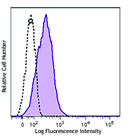
Human Fas Ligand transfected cells stained with NOK-1 PE
| Cat # | Size | Price | Quantity Check Availability | Save | ||
|---|---|---|---|---|---|---|
| 306406 | 25 tests | 121 CHF | ||||
| 306407 | 100 tests | 257 CHF | ||||
CD178 is a 38-42 kD type II glycoprotein also known as Fas ligand and CD95L. CD178 belongs to the TNF superfamily and is expressed on activated T lymphocytes, NK cells, monocytes, and granulocytes. CD178 is also expressed on parenchymal cells of the retina and cornea, retinal pigment epithelial cells, and testis. The extracellular region of FasL can be cleaved by matrix metalloproteinases (MMPs) to give rise to a 26 kD soluble protein. CD178 binds to CD95, a member of the TNFR superfamily, to induce apoptosis. CD95/CD95L interactions play an important role in the maintenance of peripheral tolerance and survival.
Product DetailsProduct Details
- Verified Reactivity
- Human
- Reported Reactivity
- Baboon
- Antibody Type
- Monoclonal
- Host Species
- Mouse
- Immunogen
- L5178Y mouse T lymphoma cells expressing recombinant human FasL
- Formulation
- Phosphate-buffered solution, pH 7.2, containing 0.09% sodium azide and BSA (origin USA)
- Preparation
- The antibody was purified by affinity chromatography, and conjugated with PE under optimal conditions.
- Concentration
- Lot-specific (to obtain lot-specific concentration and expiration, please enter the lot number in our Certificate of Analysis online tool.)
- Storage & Handling
- The antibody solution should be stored undiluted between 2°C and 8°C, and protected from prolonged exposure to light. Do not freeze.
- Application
-
FC - Quality tested
- Recommended Usage
-
Each lot of this antibody is quality control tested by immunofluorescent staining with flow cytometric analysis. For flow cytometric staining, the suggested use of this reagent is 5 µl per million cells in 100 µl staining volume or 5 µl per 100 µl of whole blood.
- Excitation Laser
-
Blue Laser (488 nm)
Green Laser (532 nm)/Yellow-Green Laser (561 nm)
- Application Notes
-
Additional reported applications (for the relevant formats) include: immunoprecipitation1,2, immunofluorescence microscopy3, immunocytochemistry2, blocking of Fas induced apoptosis1, and Western blotting11. Fas Ligand is expressed at low density on activated cells. For most successful immunofluorescent staining results, it may be important to maximize signal over background by using a relatively bright fluorochrome-antibody conjugate (Cat. No. 306407) or by using a high sensitivity, three-layer staining technique (e.g., including a biotinylated antibody (Cat. No. 306404) or biotinylated anti-mouse IgG second step (Cat. No. 405303), followed by SAv-PE (Cat. No. 405204)). In addition, applying matrix metalloproteinases (MMPS) inhibitor in the cell culture system will increase the FasL staining intensity. The Ultra-LEAF™ purified antibody (Endotoxin < 0.01 EU/µg, Azide-Free, 0.2 µm filtered) is recommended for functional assays (Cat. No. 306415 and 306416).
-
Application References
(PubMed link indicates BioLegend citation) -
- Kayagaki N, et al. 1995. J. Exp. Med. 182:1777.
- Herr I, et al. 2000. Cell Death Differ. 7:129. (WB)
- Bossi G, et al. 1999. Nature Medicine 5:90.
- Andreola G, et al. 2002. J. Exp. Med. 195:1303.
- Strauss L, et al. 2009. J. Immunol. 182:1469. PubMed
- Li JH, et al. 2009. Am J. Pathol. 175:1124. PubMed
- Zhao Q, et al. 2011. Fitoterapia. 82:735. PubMed
- Kruger K, et al. 2011. J. Appl. Physiol. 110:1226. PubMed
- Qin G, et al. 2012. J. Infect. Dis. PubMed
- Khalid M, et al. 2012. J. Virol. 86:4906. PubMed
- Qin G, et al. 2012. J. Infect. Dis. 205:1646. PubMed
- Shrestha B, et al. 2012. J. Virol. 86:8937. PubMed
- Mooren FC, et al. 2012. J. Appl. Physiol.113:1082. PubMed
- Robinet P, et al. 2014. J Immunol. 192:5332. PubMed
- Wang Y, et al. 2014. J Endocrinol. 222:151. PubMed
- Product Citations
-
- RRID
-
AB_314603 (BioLegend Cat. No. 306406)
AB_2100664 (BioLegend Cat. No. 306407)
Antigen Details
- Structure
- TNF superfamily, type II glycoprotein, 38-42 kD, 26 kD soluble form
- Distribution
-
Activated T cells, NK cells, testis, eye, neutrophils, Clara type II cells
- Function
- Apoptosis, immune privilege
- Ligand/Receptor
- CD95
- Cell Type
- Neutrophils, NK cells, T cells
- Biology Area
- Apoptosis/Tumor Suppressors/Cell Death, Cell Biology, Immunology, Neuroscience
- Molecular Family
- CD Molecules
- Antigen References
-
1. Suda T, et al. 1997. J. Exp. Med. 12:204.
2. Kayagaki N, et al. 1995. J. Exp. Med. 182:1777.
3. Tanaka M, et al. 1995. EMBO J. 14:1129. - Gene ID
- 356 View all products for this Gene ID
- UniProt
- View information about CD178 on UniProt.org
Related Pages & Pathways
Pathways


Related FAQs
- What type of PE do you use in your conjugates?
- We use R-PE in our conjugates.
Other Formats
View All CD178 Reagents Request Custom Conjugation| Description | Clone | Applications |
|---|---|---|
| Biotin anti-human CD178 (Fas-L) | NOK-1 | FC |
| PE anti-human CD178 (Fas-L) | NOK-1 | FC |
| Purified anti-human CD178 (Fas-L) | NOK-1 | FC,ICC,IP,WB |
| Brilliant Violet 421™ anti-human CD178 (Fas-L) | NOK-1 | FC |
| TotalSeq™-A0177 anti-human CD178 (Fas-L) | NOK-1 | PG |
| Ultra-LEAF™ Purified anti-human CD178 (Fas-L) | NOK-1 | FC,ICC,IP,WB |
| PE/Cyanine7 anti-human CD178 (Fas-L) | NOK-1 | FC |
| TotalSeq™-C0177 anti-human CD178 (Fas-L) Antibody | NOK-1 | PG |
| APC anti-human CD178 (Fas-L) | NOK-1 | FC |
| TotalSeq™-B0177 anti-human CD178 (Fas-L) | NOK-1 | PG |
Customers Also Purchased

Compare Data Across All Formats
This data display is provided for general comparisons between formats.
Your actual data may vary due to variations in samples, target cells, instruments and their settings, staining conditions, and other factors.
If you need assistance with selecting the best format contact our expert technical support team.
-
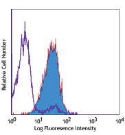
Biotin anti-human CD178 (Fas-L)

Human Fas Ligand transfected cells stained with biotinylated... -
PE anti-human CD178 (Fas-L)

Human Fas Ligand transfected cells stained with NOK-1 PE -
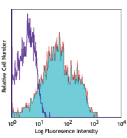
Purified anti-human CD178 (Fas-L)

Human Fas Ligand transfected cells stained with purified NOK... -
Brilliant Violet 421™ anti-human CD178 (Fas-L)

Human Fas Ligand transfected cells were stained with CD178 (... -
TotalSeq™-A0177 anti-human CD178 (Fas-L)
-
Ultra-LEAF™ Purified anti-human CD178 (Fas-L)

Human Fas Ligand transfected cells stained with LEAF™ purifi... -
PE/Cyanine7 anti-human CD178 (Fas-L)

Human Fas Ligand transfected cells were stained with CD178 (... -
TotalSeq™-C0177 anti-human CD178 (Fas-L) Antibody
-
APC anti-human CD178 (Fas-L)

Human Fas Ligand transfected cells were stained with CD178 (... -
TotalSeq™-B0177 anti-human CD178 (Fas-L)


Follow Us