 Login / Register


- Clone
- 2M2 (See other available formats)
- Regulatory Status
- RUO
- Other Names
- β2M, β2-M, beta2-microglobulin b2-M, b2M
- Isotype
- Mouse IgG1, κ
- Ave. Rating
- Submit a Review
- Product Citations
- publications

-

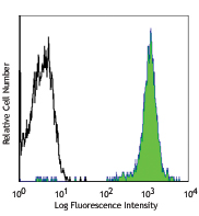
Human peripheral blood lymphocytes stained with 2M2 PE
| Cat # | Size | Price | Quantity Check Availability | Save | ||
|---|---|---|---|---|---|---|
| 316305 | 25 tests | 122 CHF | ||||
| 316306 | 100 tests | 268 CHF | ||||
β2-microglobulin (β2M) is a 12 kD nonpolymorphic Ig like protein. It is a non-membrane-anchored glycoprotein and is noncovalently associated with 39-44 kD polymorphic heavy chains of MHC class I molecules to form HLA class I antigen complex. In association with HLA class I, β2M is expressed on all leukocytes, platelets, endothelial cells, and epithelial cells. β2M plays an essential role both in governing MHC class I molecules stability and in promoting antigen binding and presenting the antigen to CD3/TCR complex of CD8+ T cells.
Product DetailsProduct Details
- Verified Reactivity
- Human
- Reported Reactivity
- African Green, Baboon, Cynomolgus, Rhesus, Pig
- Antibody Type
- Monoclonal
- Host Species
- Mouse
- Immunogen
- Purified human β2-microglobulin
- Formulation
- Phosphate-buffered solution, pH 7.2, containing 0.09% sodium azide and BSA (origin USA)
- Preparation
- The antibody was purified by affinity chromatography, and conjugated with PE under optimal conditions.
- Concentration
- Lot-specific (to obtain lot-specific concentration and expiration, please enter the lot number in our Certificate of Analysis online tool.)
- Storage & Handling
- The antibody solution should be stored undiluted between 2°C and 8°C, and protected from prolonged exposure to light. Do not freeze.
- Application
-
FC - Quality tested
- Recommended Usage
-
Each lot of this antibody is quality control tested by immunofluorescent staining with flow cytometric analysis. For flow cytometric staining, the suggested use of this reagent is 5 µl per million cells in 100 µl staining volume or 5 µl per 100 µl of whole blood.
- Excitation Laser
-
Blue Laser (488 nm)
Green Laser (532 nm)/Yellow-Green Laser (561 nm)
- Application Notes
-
Additional reported applications (for the relevant formats) include: Western blotting, and ELISA.
-
Application References
(PubMed link indicates BioLegend citation) - Product Citations
-
- RRID
-
AB_492840 (BioLegend Cat. No. 316305)
AB_492839 (BioLegend Cat. No. 316306)
Antigen Details
- Structure
- Nonpolymorphic Ig like structure, noncovalently associated with heavy chain of MHC class I molecules, 12 kD
- Distribution
-
Leukocytes, platelets, endothelial cells, epithelial cells
- Function
- Govern MHC class I molecule stability, promote MHC class I molecules binding and presenting peptide antigens to CD8+ T cells
- Ligand/Receptor
- CD3/TCR complex, CD8
- Cell Type
- Endothelial cells, Epithelial cells, Leukocytes, Platelets
- Biology Area
- Immunology
- Molecular Family
- MHC Antigens
- Antigen References
-
1. Engelhard VH. 1994. Curr. Opin. Immunol. 6:13.
2. Williams DB, et al. 1989. J. Immunol. 142:2796.
3. Danliczyk UG and TL. Delovitch. 1994. J. Immunol. 153:3533.
4. Williams A, et al. 2002. Tissue Antigens 59:3. - Gene ID
- 567 View all products for this Gene ID
- UniProt
- View information about beta2-microglobulin on UniProt.org


Related FAQs
- What type of PE do you use in your conjugates?
- We use R-PE in our conjugates.
Other Formats
View All beta2-microglobulin Reagents Request Custom Conjugation| Description | Clone | Applications |
|---|---|---|
| Purified anti-human β2-microglobulin | 2M2 | FC,WB,ELISA |
| FITC anti-human β2-microglobulin | 2M2 | FC |
| PE anti-human β2-microglobulin | 2M2 | FC |
| Biotin anti-human β2-microglobulin | 2M2 | FC |
| APC anti-human β2-microglobulin | 2M2 | FC |
| HRP anti-human β2-microglobulin | 2M2 | ELISA Detection,FC |
| PE/Cyanine7 anti-human β2-microglobulin | 2M2 | FC |
| APC/Fire™ 750 anti-human β2-microglobulin | 2M2 | FC |
| PerCP/Cyanine5.5 anti-human β2-microglobulin | 2M2 | FC |
| PE/Dazzle™ 594 anti-human β2-microglobulin | 2M2 | FC |
| TotalSeq™-A0057 anti-human β2-microglobulin | 2M2 | PG |
| TotalSeq™-C0057 anti-human β2-microglobulin | 2M2 | PG |
| TotalSeq™-B0057 anti-human β2-microglobulin | 2M2 | PG |
Customers Also Purchased
Compare Data Across All Formats
This data display is provided for general comparisons between formats.
Your actual data may vary due to variations in samples, target cells, instruments and their settings, staining conditions, and other factors.
If you need assistance with selecting the best format contact our expert technical support team.
-
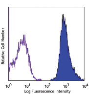
Purified anti-human β2-microglobulin

Human peripheral blood lymphocytes stained with purified 2M2... -
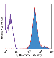
FITC anti-human β2-microglobulin

Human peripheral blood lymphocytes stained with 2M2 FITC -
PE anti-human β2-microglobulin

Human peripheral blood lymphocytes stained with 2M2 PE -
Biotin anti-human β2-microglobulin

Human peripheral blood lymphocytes stained with biotinylated... -
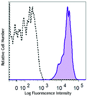
APC anti-human β2-microglobulin

Human peripheral blood lymphocytes stained with 2M2 APC -
HRP anti-human β2-microglobulin
-
PE/Cyanine7 anti-human β2-microglobulin

Human peripheral blood lymphocytes were stained with anti-hu... -
APC/Fire™ 750 anti-human β2-microglobulin

Human peripheral blood lymphocytes were stained with anti-hu... -
PerCP/Cyanine5.5 anti-human β2-microglobulin

Human peripheral blood lymphocytes were stained with ß2-micr... -
PE/Dazzle™ 594 anti-human β2-microglobulin

Human peripheral blood lymphocytes were stained with anti-hu... -
TotalSeq™-A0057 anti-human β2-microglobulin
-
TotalSeq™-C0057 anti-human β2-microglobulin
-
TotalSeq™-B0057 anti-human β2-microglobulin


Follow Us