 Login / Register


- Clone
- TS2/7 (See other available formats)
- Regulatory Status
- RUO
- Other Names
- α1 integrin, VLA-1 α chain, Integrin α1 chain, ITGA1
- Isotype
- Mouse IgG1, κ
- Ave. Rating
- Submit a Review
- Product Citations
- publications

-

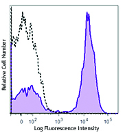
Human cervical cancer cell line, Hela, stained with TS2/7 FITC
| Cat # | Size | Price | Quantity Check Availability | Save | ||
|---|---|---|---|---|---|---|
| 328307 | 25 tests | 94 CHF | ||||
| 328308 | 100 tests | 211 CHF | ||||
CD49a is a 200 kD type I transmembrane glycoprotein also known as α1 integrin, VLA-1 α chain, or Integrin α1. It associates with CD29 (β1 integrin) to form VLA-1 complex, a collagen IV and alminin-1 receptor. It is expressed on activated T cells, monocytes, NK cells, smooth muscle cells, neuronal cells, fibroblasts, and mesenchymal cells. CD49a is an adhesion molecule and is involved in the regulation of leukocyte migration, T cell proliferation, and cytokine production.
Product DetailsProduct Details
- Verified Reactivity
- Human
- Reported Reactivity
- African Green, Baboon, Cynomolgus, Rhesus
- Antibody Type
- Monoclonal
- Host Species
- Mouse
- Immunogen
- Human CTL line
- Formulation
- Phosphate-buffered solution, pH 7.2, containing 0.09% sodium azide and BSA (origin USA)
- Preparation
- The antibody was purified by affinity chromatography, and conjugated with FITC under optimal conditions.
- Concentration
- Lot-specific (to obtain lot-specific concentration and expiration, please enter the lot number in our Certificate of Analysis online tool.)
- Storage & Handling
- The antibody solution should be stored undiluted between 2°C and 8°C, and protected from prolonged exposure to light. Do not freeze.
- Application
-
FC - Quality tested
- Recommended Usage
-
Each lot of this antibody is quality control tested by immunofluorescent staining with flow cytometric analysis. For flow cytometric staining, the suggested use of this reagent is 5 µl per million cells in 100 µl staining volume or 5 µl per 100 µl of whole blood.
- Excitation Laser
-
Blue Laser (488 nm)
- Application Notes
-
Additional reported applications include: immunoprecipitation1, immunohistochemical staining1 of acetone-fixed frozen tissue sections, and spatial biology (IBEX)3,4.
-
Application References
(PubMed link indicates BioLegend citation) - Product Citations
-
- RRID
-
AB_1236430 (BioLegend Cat. No. 328307)
AB_2129084 (BioLegend Cat. No. 328308)
Antigen Details
- Structure
- Integrin alpha chain family, Type I membrane protein, alpha chain of heterodimeric integrin receptor, 200 kD. Associates with CD29 to form VLA-1 complex
- Distribution
-
Activated T cells, monocytes, NK cells, smooth muscle cells, neuronal cells, fibroblasts, and mesenchymal cells
- Function
- Adhesion, leukocyte migration
- Ligand/Receptor
- With integrin β1 (CD29) forms receptor for laminin-1 and collagen IV
- Cell Type
- Fibroblasts, Mesenchymal cells, Mesenchymal Stem Cells, Monocytes, NK cells, T cells
- Biology Area
- Immunology, Stem Cells
- Molecular Family
- Adhesion Molecules, CD Molecules
- Antigen References
-
- Zola H, et al. Eds. 2007. Leukocyte and Stromal Cell Molecules:The CD Markers. Wiley-Liss Press. p122
- Boiret N, et al. 2005. Exp. Hematol. 33:219
- Gene ID
- 3672 View all products for this Gene ID
- UniProt
- View information about CD49a on UniProt.org
Related Pages & Pathways
Pathways


Related FAQs
Other Formats
View All CD49a Reagents Request Custom Conjugation| Description | Clone | Applications |
|---|---|---|
| Purified anti-human CD49a | TS2/7 | FC |
| FITC anti-human CD49a | TS2/7 | FC |
| PE anti-human CD49a | TS2/7 | FC,SB |
| Alexa Fluor® 647 anti-human CD49a | TS2/7 | FC,SB |
| PE/Cyanine7 anti-human CD49a | TS2/7 | FC |
| APC anti-human CD49a | TS2/7 | FC |
| TotalSeq™-A0575 anti-human CD49a | TS2/7 | PG |
| APC/Fire™ 750 anti-human CD49a | TS2/7 | FC |
| TotalSeq™-C0575 anti-human CD49a | TS2/7 | PG |
| PerCP/Cyanine5.5 anti-human CD49a | TS2/7 | FC |
| TotalSeq™-B0575 anti-human CD49a Antibody | TS2/7 | PG |
Customers Also Purchased
Compare Data Across All Formats
This data display is provided for general comparisons between formats.
Your actual data may vary due to variations in samples, target cells, instruments and their settings, staining conditions, and other factors.
If you need assistance with selecting the best format contact our expert technical support team.
-
Purified anti-human CD49a

Human cervical cancer cell line, Hela, stained with purified... -
FITC anti-human CD49a

Human cervical cancer cell line, Hela, stained with TS2/7 FI... -
PE anti-human CD49a

Human cervical cancer cell line, Hela, stained with TS2/7 PE 
Confocal image of human liver sample acquired using the IBEX... -
Alexa Fluor® 647 anti-human CD49a

Human cervical cancer cell line, Hela, stained with TS2/7 Al... 
Confocal image of human spleen sample acquired using the IBE... 
Confocal image of human lymph node sample acquired using the... 
Confocal image of human jejunum sample acquired using the IB... -
PE/Cyanine7 anti-human CD49a

HeLa cells (Human cervical cancer cell lines) were stained w... -
APC anti-human CD49a

HeLa cells (Human cervical cancer cell lines) were stained w... -
TotalSeq™-A0575 anti-human CD49a
-
APC/Fire™ 750 anti-human CD49a

Human cervical cancer cell line, HeLa, was stained with CD49... -
TotalSeq™-C0575 anti-human CD49a
-
PerCP/Cyanine5.5 anti-human CD49a

HeLa cells (human cervical cancer cell lines) were stained w... -
TotalSeq™-B0575 anti-human CD49a Antibody


Follow Us