 Login / Register


- Clone
- 281-2 (See other available formats)
- Regulatory Status
- RUO
- Other Names
- Syndecan-1
- Isotype
- Rat IgG2a, κ
- Ave. Rating
- Submit a Review
- Product Citations
- publications

-

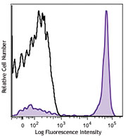
C57BL/6 mouse bone marrow cells were stained with CD19 APC and CD138 (clone 281-2) Brilliant Violet 605™ (top) or rat IgG2a, κ Brilliant Violet 605™ isotype control (bottom). -

CD138, a member of the syndecan protein family, is a type I integral membrane heparin sulfate proteoglycan also known as Syndecan-1. Syndecan-1 participates in cell proliferation, cell migration, and cell matrix adhesion via interaction with collagen, fibronectin, and other soluble molecules (such as FGF-basic). It is expressed on normal and malignant plasma cells, pre-B cells, mesenchymal cells, epithelial cells, and endothelial cells.
Product DetailsProduct Details
- Verified Reactivity
- Mouse
- Antibody Type
- Monoclonal
- Host Species
- Rat
- Immunogen
- Mouse mammary gland epithelial cell line NMuMG
- Formulation
- Phosphate-buffered solution, pH 7.2, containing 0.09% sodium azide and BSA (origin USA).
- Preparation
- The antibody was purified by affinity chromatography and conjugated with Brilliant Violet 605™ under optimal conditions.
- Concentration
- µg sizes: 0.2 mg/mLµL sizes: lot-specific (to obtain lot-specific concentration and expiration, please enter the lot number in our Certificate of Analysis online tool.)
- Storage & Handling
- The antibody solution should be stored undiluted between 2°C and 8°C, and protected from prolonged exposure to light. Do not freeze.
- Application
-
FC - Quality tested
- Recommended Usage
-
Each lot of this antibody is quality control tested by immunofluorescent staining with flow cytometric analysis. For flow cytometric staining using the µl size, the suggested use of this reagent is 5 µl per million cells in 100 µl staining volume or 5 µl per 100 µl of whole blood. For flow cytometric staining using the µg size, the suggested use of this reagent is ≤0.5 µg per million cells in 100 µl volume. It is recommended that the reagent be titrated for optimal performance for each application.
Brilliant Violet 605™ excites at 405 nm and emits at 603 nm. The bandpass filter 610/20 nm is recommended for detection, although filter optimization may be required depending on other fluorophores used. Be sure to verify that your cytometer configuration and software setup are appropriate for detecting this channel. Refer to your instrument manual or manufacturer for support. Brilliant Violet 605™ is a trademark of Sirigen Group Ltd.
Learn more about Brilliant Violet™.
This product is subject to proprietary rights of Sirigen Inc. and is made and sold under license from Sirigen Inc. The purchase of this product conveys to the buyer a non-transferable right to use the purchased product for research purposes only. This product may not be resold or incorporated in any manner into another product for resale. Any use for therapeutics or diagnostics is strictly prohibited. This product is covered by U.S. Patent(s), pending patent applications and foreign equivalents. - Excitation Laser
-
Violet Laser (405 nm)
- Application Notes
-
Additional reported applications (for the relevant formats) include: immunohistochemical staining of frozen tissue3 and formalin-fixed paraffin embedded tissue4 and immunofluorescent staining2,3.
-
Application References
(PubMed link indicates BioLegend citation) -
- Jalkanen M, et al. 1985. J. Cell. Biol. 101:976. (FC)
- Miettinen H, et al. 1994. J. Cell. Sci. 107:1571. (IF)
- Li Q, et al. 2002. Cell 111:635. (IF, IHC)
- McCarthy BA, et al. 2012. BMC Cancer. 12:203. (IHC)
- Product Citations
-
- RRID
-
AB_2562336 (BioLegend Cat. No. 142515)
AB_2715767 (BioLegend Cat. No. 142531)
AB_2562337 (BioLegend Cat. No. 142516)
Antigen Details
- Structure
- Transmembrane heparan sulfate proteoglycan; member of the syndecan proteoglycan family, 100-200 kD
- Function
- Cell adhesion, cell motility, cell migration, cell proliferation
- Cell Type
- B cells
- Biology Area
- Apoptosis/Tumor Suppressors/Cell Death, Cell Adhesion, Cell Biology, Cell Motility/Cytoskeleton/Structure, Immunology
- Molecular Family
- Adhesion Molecules, CD Molecules
- Antigen References
-
1. Zong F, et al. 2011. PLoS ONE 6:e14816.
2. Yamashita Y, et al. 1999. J. Immunol. 162:5940.
3. Sanderson RD, et al. 1989. Cell. Regul. 1:27. - Gene ID
- 20969 View all products for this Gene ID
- UniProt
- View information about CD138 on UniProt.org
Related Pages & Pathways
Pathways


Related FAQs
Other Formats
View All CD138 Reagents Request Custom ConjugationCustomers Also Purchased
Compare Data Across All Formats
This data display is provided for general comparisons between formats.
Your actual data may vary due to variations in samples, target cells, instruments and their settings, staining conditions, and other factors.
If you need assistance with selecting the best format contact our expert technical support team.
-
Purified anti-mouse CD138 (Syndecan-1)

C57BL/6 mouse bone marrow cells were stained with CD19 FITC ... 
-
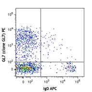
PE anti-mouse CD138 (Syndecan-1)
C57BL/6 mouse bone marrow cells were stained with CD19 FITC ...
-
APC anti-mouse CD138 (Syndecan-1)

C57BL/6 mouse bone marrow cells were stained with CD19 FITC ... 
-
Brilliant Violet 421™ anti-mouse CD138 (Syndecan-1)

C57BL/6 mouse bone marrow cells were stained with CD19 APC a... 
-
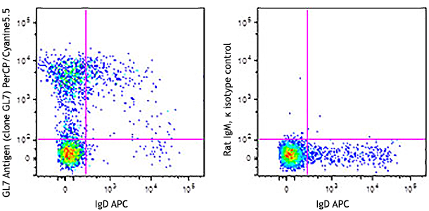
PerCP/Cyanine5.5 anti-mouse CD138 (Syndecan-1)

C57BL/6 mouse bone marrow cells were stained with CD19 FITC ... -
Biotin anti-mouse CD138 (Syndecan-1)

C57BL/6 mouse splenocytes were stained with CD19 APC and bio... 
-
PE/Cyanine7 anti-mouse CD138 (Syndecan-1)

C57BL/6 mouse splenocytes were stained with CD19 APC and CD1... 
-
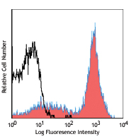
Brilliant Violet 605™ anti-mouse CD138 (Syndecan-1)

C57BL/6 mouse bone marrow cells were stained with CD19 APC a... 
-
Brilliant Violet 650™ anti-mouse CD138 (Syndecan-1)

C57BL/6 mouse bone marrow cells were stained with CD19 FITC ... 
-
Brilliant Violet 711™ anti-mouse CD138 (Syndecan-1)

C57BL/6 mouse splenocytes were stained with CD19 APC and CD1... -
Brilliant Violet 510™ anti-mouse CD138 (Syndecan-1)

C57BL/6 mouse bone marrow cells were stained with CD19 APC a... 
-
Alexa Fluor® 647 anti-mouse CD138 (Syndecan-1)

C57BL/6 mouse bone marrow cells were stained with CD19 FITC ... 
-
PE/Dazzle™ 594 anti-mouse CD138 (Syndecan-1)

C57BL/6 mouse bone marrow cells were stained with CD19 APC a... 
-
APC/Cyanine7 anti-mouse CD138 (Syndecan-1)

C57BL/6 mouse bone marrow cells were stained with CD19 FITC ... -
TotalSeq™-A0810 anti-mouse CD138 (Syndecan-1)
-
Brilliant Violet 785™ anti-mouse CD138 (Syndecan-1)

C57BL/6 mouse bone marrow cells were stained with CD19 FITC ... -
TotalSeq™-C0810 anti-mouse CD138 (Syndecan-1)
-
TotalSeq™-B0810 anti-mouse CD138 (Syndecan-1)
-
PE/Cyanine5 anti-mouse CD138 (Syndecan-1)

C57BL/6 mouse bone marrow cells were stained with anti-mouse... -
APC/Fire™ 750 anti-mouse CD138 (Syndecan-1)

C577BL/6 mouse bone marrow cells were stained with anti-mous... -
PE/Fire™ 640 anti-mouse CD138 (Syndecan-1)

C57BL/6 mouse bone marrow cells were stained with anti-mouse... -
PE/Fire™ 810 anti-mouse CD138 (Syndecan-1)

C57BL/6 mouse bone marrow cells were stained with anti-mouse...


Follow Us