 Login / Register


- Clone
- O323 (See other available formats)
- Regulatory Status
- RUO
- Workshop
- IV T-186
- Other Names
- S152, T14, TNFRSF7
- Isotype
- Mouse IgG1, κ
- Ave. Rating
- Submit a Review
- Product Citations
- publications

-

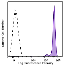
Human peripheral blood lymphocytes were stained with CD27 (clone O323) Brilliant Violet 510™.
| Cat # | Size | Price | Quantity Check Availability | Save | ||
|---|---|---|---|---|---|---|
| 302835 | 25 tests | 193 CHF | ||||
| 302836 | 100 tests | 402 CHF | ||||
CD27 is a 50-55 kD type I membrane protein also known as S152 and T14. It is a lymphocyte-specific member of the TNF-receptor superfamily. CD27 is expressed on medullary thymocytes, virtually all mature T cells, some B cells, and NK cells. CD27 binds to CD70 and plays an important role in costimulation of T cell activation, and regulation of B cell differentiation and proliferation. The cytoplasmic domains of CD27 have also been shown to interact with TRAF2 and TRAF5 to elicit NF-κB and SAPK/JNK activation.
Product DetailsProduct Details
- Verified Reactivity
- Human, Cynomolgus, Rhesus
- Reported Reactivity
- African Green, Baboon, Squirrel Monkey
- Antibody Type
- Monoclonal
- Host Species
- Mouse
- Formulation
- Phosphate-buffered solution, pH 7.2, containing 0.09% sodium azide and BSA (origin USA).
- Preparation
- The antibody was purified by affinity chromatography and conjugated with Brilliant Violet 510™ under optimal conditions.
- Concentration
- Lot-specific (to obtain lot-specific concentration and expiration, please enter the lot number in our Certificate of Analysis online tool.)
- Storage & Handling
- The antibody solution should be stored undiluted between 2°C and 8°C, and protected from prolonged exposure to light. Do not freeze.
- Application
-
FC - Quality tested
- Recommended Usage
-
Each lot of this antibody is quality control tested by immunofluorescent staining with flow cytometric analysis. For flow cytometric staining, the suggested use of this reagent is 5 µl per million cells in 100 µl staining volume or 5 µl per 100 µl of whole blood.
Brilliant Violet 510™ excites at 405 nm and emits at 510 nm. The bandpass filter 510/50 nm is recommended for detection, although filter optimization may be required depending on other fluorophores used. Be sure to verify that your cytometer configuration and software setup are appropriate for detecting this channel. Refer to your instrument manual or manufacturer for support. Brilliant Violet 510™ is a trademark of Sirigen Group Ltd.
Learn more about Brilliant Violet™.
This product is subject to proprietary rights of Sirigen Inc. and is made and sold under license from Sirigen Inc. The purchase of this product conveys to the buyer a non-transferable right to use the purchased product for research purposes only. This product may not be resold or incorporated in any manner into another product for resale. Any use for therapeutics or diagnostics is strictly prohibited. This product is covered by U.S. Patent(s), pending patent applications and foreign equivalents. - Excitation Laser
-
Violet Laser (405 nm)
-
Application References
(PubMed link indicates BioLegend citation) -
- Knapp W, et al. Eds. 1989. Leucocyte Typing IV. Oxford University Press. New York.
- Correia DV, et al. 2011. Blood 118:992. (FC) PubMed
- Product Citations
-
- RRID
-
AB_2561382 (BioLegend Cat. No. 302835)
AB_2562086 (BioLegend Cat. No. 302836)
Antigen Details
- Structure
- TNF-R superfamily, type I transmembrane glycoprotein, 50-55 kD
- Distribution
-
Medullary thymocytes, T and B cell subsets, NK cells
- Function
- T cell costimulation
- Ligand/Receptor
- CD70
- Cell Type
- B cells, NK cells, T cells, Thymocytes, Tregs
- Biology Area
- Costimulatory Molecules, Immunology
- Molecular Family
- CD Molecules
- Antigen References
-
1. Hintzen R, et al. 1994. Immunol. Today 15:307.
2. Agematsu K, et al. 1995. J. Immunol. 154:3627. - Gene ID
- 939 View all products for this Gene ID
- UniProt
- View information about CD27 on UniProt.org
Related Pages & Pathways
Pathways


Related FAQs
Other Formats
View All CD27 Reagents Request Custom ConjugationCustomers Also Purchased
Compare Data Across All Formats
This data display is provided for general comparisons between formats.
Your actual data may vary due to variations in samples, target cells, instruments and their settings, staining conditions, and other factors.
If you need assistance with selecting the best format contact our expert technical support team.
-
APC anti-human CD27

Human peripheral blood lymphocytes stained with O323 APC -
Biotin anti-human CD27

Human peripheral blood lymphocytes stained with biotinylated... -
FITC anti-human CD27

Human peripheral blood lymphocytes stained with O323 FITC -
PE anti-human CD27

Human peripheral blood lymphocytes stained with O323 PE 
PE anti-human CD27 Antibody Human peripheral blood was stai... -
Purified anti-human CD27

Human peripheral blood lymphocytes stained with purified O32... -
Alexa Fluor® 647 anti-human CD27

Human peripheral blood lymphocytes stained with O323 Alexa F... -
Alexa Fluor® 700 anti-human CD27

Human peripheral blood lymphocytes stained with O323 Alexa F... -
APC/Cyanine7 anti-human CD27

Human peripheral blood lymphocytes stained with CD27 (clone ... -
PerCP anti-human CD27

Human peripheral blood lymphocytes stained with O323 PerCP -
PerCP/Cyanine5.5 anti-human CD27

Human peripheral blood lymphocytes were stained with CD27 (C... -
Pacific Blue™ anti-human CD27

Human peripheral blood lymphocytes stained with O323 Pacific... -
Brilliant Violet 421™ anti-human CD27

Human peripheral blood lymphocytes were stained with CD27 (c... -
Brilliant Violet 570™ anti-human CD27

Human peripheral blood lymphocytes were stained with CD27 (c... -
Brilliant Violet 650™ anti-human CD27

Human peripheral blood lymphocytes were stained with CD27 (c... -
Brilliant Violet 605™ anti-human CD27

Human peripheral blood lymphocytes were stained with CD27 (c... -
Brilliant Violet 711™ anti-human CD27

Human peripheral blood lymphocytes were stained with CD27 (c... -
Brilliant Violet 785™ anti-human CD27

Human peripheral blood lymphocytes were stained with CD27 (c... -
Brilliant Violet 510™ anti-human CD27

Human peripheral blood lymphocytes were stained with CD27 (c... -
PE/Cyanine7 anti-human CD27

Human peripheral blood lymphocytes were stained with CD27 (c... -
Purified anti-human CD27 (Maxpar® Ready)

Human PBMCs stained with 154Sm-anti-CD45 (HI30) and 167Er-an... -
PE/Dazzle™ 594 anti-human CD27

Human peripheral blood lymphocytes were stained with CD27 (c... -
APC/Fire™ 750 anti-human CD27

Human peripheral blood lymphocytes were stained with CD27 (c... -
TotalSeq™-A0154 anti-human CD27
-
Brilliant Violet 750™ anti-human CD27

Human peripheral blood lymphocytes were stained with CD27 (c... -
TotalSeq™-B0154 anti-human CD27
-
TotalSeq™-C0154 anti-human CD27
-
Spark NIR™ 685 anti-human CD27

Human peripheral blood lymphocytes were stained with CD27 (c... -
PE/Fire™ 810 anti-human CD27

Human peripheral lymphocytes were stained with CD27 (clone O... -
TotalSeq™-D0154 anti-human CD27
-
APC/Fire™ 810 anti-human CD27

Human peripheral lymphocytes were stained with anti-human CD... -
PE/Cyanine5 anti-human CD27 Antibody

Human peripheral blood lymphocytes were stained with anti-hu... -
Spark UV™ 387 anti-human CD27

Human peripheral blood lymphocytes were stained with anti-hu... -
Spark Blue™ 515 anti-human CD27

Human peripheral blood lymphocytes were stained with anti-hu...


Follow Us