 Login / Register


- Clone
- 1F4 (See other available formats)
- Regulatory Status
- RUO
- Other Names
- T cell antigen receptor complex, T3
- Isotype
- Mouse IgM, κ
- Ave. Rating
- Submit a Review
- Product Citations
- publications

-

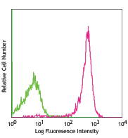
LOU rat splenocytes stained with 1F4 Alexa Fluor® 647 -

Rat frozen spleen section was fixed with 4% paraformaldehyde (PFA) for ten minutes at room temperature and blocked with 5% FBS for 30 minutes at room temperature. Then the section was stained with 10 µg/mL of CD11b/c (clone OX-42) Alexa Fluor® 488 (blue), 10 µg/mL of CD3 (clone 1F4) Alexa Fluor® 647 (yellow), and 10 µg/mL of purified CD38 (clone 14.27) overnight at 4°C, followed by 5 µg/mL of anti-mouse IgG2b (clone RMG2b-1) Alexa Fluor® 594 (red) for two hours at room temperature. The image was captured by 10X objective.
| Cat # | Size | Price | Quantity Check Availability | Save | ||
|---|---|---|---|---|---|---|
| 201408 | 100 µg | 229 CHF | ||||
CD3 is a complex composed of δ, γ, ε, and ζ chains. They are 20-25 kD members of the immunoglobulin superfamily and associated with the T cell receptor (TCR). CD3 is expressed on thymocytes, peripheral T cells, some NK-T cells, and dendritic epidermal T cells. CD3 is involved in antigen recognition, signal transduction, and T cell activation.
Product DetailsProduct Details
- Verified Reactivity
- Rat
- Antibody Type
- Monoclonal
- Host Species
- Mouse
- Immunogen
- F344 rat spleen cells stimulated with PMA and calcium ionophore
- Formulation
- Phosphate-buffered solution, pH 7.2, containing 0.09% sodium azide.
- Preparation
- The antibody was purified by affinity chromatography and conjugated with Alexa Fluor® 647 under optimal conditions.
- Concentration
- 0.5 mg/ml
- Storage & Handling
- The antibody solution should be stored undiluted between 2°C and 8°C, and protected from prolonged exposure to light. Do not freeze.
- Application
-
FC - Quality tested
IHC-F - Verified - Recommended Usage
-
Each lot of this antibody is quality control tested by immunofluorescent staining with flow cytometric analysis. For flow cytometric staining, the suggested use of this reagent is ≤0.06 µg per million cells in 100 µl volume. For immunohistochemistry staining on frozen tissue sections, a concentration range of 2.5 - 10 µg/ml is suggested. It is recommended that the reagent be titrated for optimal performance for each application.
* Alexa Fluor® 647 has a maximum emission of 668 nm when it is excited at 633 nm / 635 nm.
Alexa Fluor® and Pacific Blue™ are trademarks of Life Technologies Corporation.
View full statement regarding label licenses - Excitation Laser
-
Red Laser (633 nm)
- Application Notes
-
Immobilized 1F4 antibody can induce T cell proliferation in vitro. Additional reported applications (for relevant formats of this clone) include: immunohistochemistry of acetone-fixed frozen sections1 and formaldehyde- fixed paraffin embedded sections4,5 immunofluorescence microscopy3, in vivo activation of T cell responses1, and in vivo inhibition of T cell responses2.
-
Application References
(PubMed link indicates BioLegend citation) -
- Tanaka T, et al. 1989. J. Immunol. 142:2791. (Activ, IHC, IP)
- Nicholls MR, et al. 1993. Transplantation 55:459. (Block)
- Elbe A, et al. 1993. J. Invest. Dermatol. 102:74. (IF)
- Baba T, et al. 2006. Blood 107:2004. (IHC)
- Fujishiro J, et al. 2010. Am. J. Transplant. 10:1545-55. (IHC-P)
- Li X, et al. 2009. J. Immunol. 183:3955. (FC) PubMed
- Product Citations
-
- RRID
-
AB_893304 (BioLegend Cat. No. 201408)
Antigen Details
- Structure
- Ig superfamily, approximately 20-25 kD
- Distribution
-
Thymocytes, peripheral T cells, dendritic epidermal T cells, NK-T cells
- Function
- Antigen recognition, TCR signal transduction, T cell activation
- Ligand/Receptor
- Peptide antigen/MHC complex
- Cell Type
- NKT cells, T cells, Thymocytes
- Biology Area
- Immunology
- Molecular Family
- CD Molecules
- Antigen References
-
1. Tanaka T, et al. 1989 J. Immunol. 142:2791.
2. Elbe A, et al. 1993. J. Invest. Dermatol. 102:74. - Gene ID
- 25710 View all products for this Gene ID 300678 View all products for this Gene ID 315609 View all products for this Gene ID 25300 View all products for this Gene ID
- UniProt
- View information about CD3 on UniProt.org
Related Pages & Pathways
Pathways


Related FAQs
Other Formats
View All CD3 Reagents Request Custom Conjugation| Description | Clone | Applications |
|---|---|---|
| Purified anti-rat CD3 | 1F4 | FC,IHC-F |
| FITC anti-rat CD3 | 1F4 | FC |
| PE anti-rat CD3 | 1F4 | FC |
| Alexa Fluor® 488 anti-rat CD3 | 1F4 | FC,IHC-F |
| Alexa Fluor® 647 anti-rat CD3 | 1F4 | FC,IHC-F |
| APC anti-rat CD3 | 1F4 | FC |
| Ultra-LEAF™ Purified anti-rat CD3 | 1F4 | FC,IHC-F |
| PerCP/Cyanine5.5 anti-rat CD3 | 1F4 | FC |
| PE/Cyanine5 anti-rat CD3 | 1F4 | FC |
| PE/Cyanine7 anti-rat CD3 | 1F4 | FC |
| Spark Violet™ 423 anti-rat CD3 | 1F4 | FC |
Customers Also Purchased
Compare Data Across All Formats
This data display is provided for general comparisons between formats.
Your actual data may vary due to variations in samples, target cells, instruments and their settings, staining conditions, and other factors.
If you need assistance with selecting the best format contact our expert technical support team.
-
Purified anti-rat CD3

LOU rat splenocytes stained with purified 1F4, followed by a... -
FITC anti-rat CD3

LOU rat splenocytes stained with 1F4 FITC -
PE anti-rat CD3

Lewis rat splenocytes were stained with CD3 (clone 1F4) PE (... -
Alexa Fluor® 488 anti-rat CD3

LOU rat splenocytes stained with 1F4 Alexa Fluor® 488 
Rat frozen spleen section was fixed with 4% paraformaldehyde... -
Alexa Fluor® 647 anti-rat CD3

LOU rat splenocytes stained with 1F4 Alexa Fluor® 647 
Rat frozen spleen section was fixed with 4% paraformaldehyde... -
APC anti-rat CD3

Lewis rat splenocytes were stained with CD3 (clone 1F4) APC ... -
Ultra-LEAF™ Purified anti-rat CD3
-
PerCP/Cyanine5.5 anti-rat CD3

Lewis rat splenocytes were stained with CD3 (clone 1F4) Per... -
PE/Cyanine5 anti-rat CD3

Lewis rat splenocytes were stained with anti-rat CD3 (clone ... -
PE/Cyanine7 anti-rat CD3

Lewis rat splenocytes were stained with anti-rat CD3 (clone ... -
Spark Violet™ 423 anti-rat CD3

Lewis rat splenocytes were stained with anti-rat CD3 (clone ...


Follow Us