 Login / Register


- Clone
- 4B11B69 (See other available formats)
- Regulatory Status
- RUO
- Other Names
- P44/42 MAP (motogen activated protein) Kinase, Extracelluar signal-regulated kinase (ERK)
- Isotype
- Mouse IgG2b, κ
- Ave. Rating
- Submit a Review
- Product Citations
- publications

-

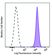
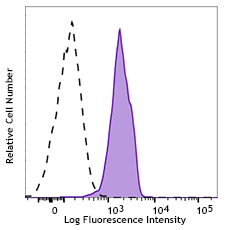
Human peripheral blood lymphocytes were stimulated with (filled histogram) or without (open histogram) Cell Activation Cocktail (without Brefeldin A) for 15 minutes, then fixed with Fixation Buffer, permeabilized with True-Phos™ Perm Buffer, and intracellularly stained with ERK1/2 Phospho (Thr202/Tyr204) (clone 4B11B69) Alexa Fluor® 488.
| Cat # | Size | Price | Quantity Check Availability | Save | ||
|---|---|---|---|---|---|---|
| 675507 | 25 tests | 147 CHF | ||||
| 675508 | 100 tests | 358 CHF | ||||
ERK1/2 are members of mitogen-activated kinases (MAPKs) of serine/threonine protein kinases. ERK1/2 can be activated by a range of extracellular stimuli, such as mitogen, growth factors, neurotransmitters, chemokines, and cytokines, through receptor tyrosine kinases (RTK), G protein-coupled receptors (GPCRs), or protein kinase C (PKC). Upon stimulation, ERK1/2 are phosphorylated by the upstream kinase MEK on residues Thr202 and Tyr204 and in turn phosphorylate many other downstream molecules that are involved in a range of cellular processes such as cell proliferation, differentiation, motility and cell death.
Product DetailsProduct Details
- Verified Reactivity
- Human, Mouse
- Antibody Type
- Monoclonal
- Host Species
- Mouse
- Immunogen
- Synthetic peptide (TGFLT*EY*VATRC) conjugated to KLH.
- Formulation
- Phosphate-buffered solution, pH 7.2, containing 0.09% sodium azide and BSA (origin USA)
- Preparation
- The antibody was purified by affinity chromatography and conjugated with Alexa Fluor® 488 under optimal conditions.
- Concentration
- Lot-specific (to obtain lot-specific concentration and expiration, please enter the lot number in our Certificate of Analysis online tool.)
- Storage & Handling
- The antibody solution should be stored undiluted between 2°C and 8°C, and protected from prolonged exposure to light. Do not freeze.
- Application
-
ICFC - Quality tested
- Recommended Usage
-
Each lot of this antibody is quality control tested by intracellular flow cytometry using our True-Phos™ Perm Buffer in Cell Suspensions Protocol. For flow cytometric staining, the suggested use of this reagent is 5 µl per million cells in 100 µl staining volume or 5 µl per 100 µl of whole blood.
* Alexa Fluor® 488 has a maximum emission of 519 nm when it is excited at 488 nm.
Alexa Fluor® and Pacific Blue™ are trademarks of Life Technologies Corporation.
View full statement regarding label licenses - Excitation Laser
-
Blue Laser (488 nm)
- Product Citations
-
- RRID
-
AB_2629783 (BioLegend Cat. No. 675507)
AB_2629784 (BioLegend Cat. No. 675508)
Antigen Details
- Structure
- ERK1 and ERK2 encode 42 and 44 kD protein kinases and share 85% homology.
- Distribution
-
Cytoplasm. Nucleus.
- Function
- ERK1/2 are broadly expressed protein kinases that are involved in proliferation, differentiation, motility, and cell death. In response to extracellular stimuli, ERK1/2 are phosphorylated at Thr202 and Tyr204 and in turn transduce signals to downstream substrates.
- Interaction
- ERK1/2 phosphorylates a wide range of downstream substrates, such as ATF2, BCL6, EIF4EBP1, CANX, MAP2, DAPK1, RAF1, SYK, and GRB10.
- Biology Area
- Cell Biology, Neuroscience, Signal Transduction, Synaptic Biology
- Molecular Family
- Postsynaptic proteins, Phospho-Proteins, Protein Kinases/Phosphatase
- Antigen References
-
1. Futran AS, et al. 2013. Curr. Biol. 23:R972.
2. Mendoza MC, et al. 2011. Trends Biochem. Sci. 36:320.
3. Chambard JC, et al. 2007. Biochim. Biophys. Acta. 1773:1299.
4. Roux PP, et al. 2004. Microbiol. Mol. Biol. Rev. 68:320.
5. Torii S, et al. 2004. J. Biochem. 136:557.
6. Clark EA, et al. 1996. J. Biol. Chem. 271:14814. - Gene ID
- 5594 View all products for this Gene ID
- UniProt
- View information about ERK1/2 on UniProt.org


Related FAQs
Other Formats
View All ERK1/2 Reagents Request Custom Conjugation| Description | Clone | Applications |
|---|---|---|
| Purified anti-ERK1/2 Phospho (Thr202/Tyr204) | 4B11B69 | WB,ICC |
| Direct-Blot™ HRP anti-ERK1/2 Phospho (Thr202/Tyr204) | 4B11B69 | WB |
| Alexa Fluor® 647 anti-ERK1/2 Phospho (Thr202/Tyr204) | 4B11B69 | ICFC |
| Alexa Fluor® 488 anti-ERK1/2 Phospho (Thr202/Tyr204) | 4B11B69 | ICFC |
Customers Also Purchased
Compare Data Across All Formats
This data display is provided for general comparisons between formats.
Your actual data may vary due to variations in samples, target cells, instruments and their settings, staining conditions, and other factors.
If you need assistance with selecting the best format contact our expert technical support team.
-
Purified anti-ERK1/2 Phospho (Thr202/Tyr204)

Total cell lysates (15 µg protein) from Jurkat cells without... 
Untreated (left) and PMA-treated (20 nM PMA treatment for 10... -
Direct-Blot™ HRP anti-ERK1/2 Phospho (Thr202/Tyr204)

15 µg of total protein extract from Jurkat cells treated wit... -
Alexa Fluor® 647 anti-ERK1/2 Phospho (Thr202/Tyr204)

Human peripheral blood lymphocytes were stimulated with (fil... 
C57BL/6 mouse splenocytes were stimulated with (filled histo... -
Alexa Fluor® 488 anti-ERK1/2 Phospho (Thr202/Tyr204)

Human peripheral blood lymphocytes were stimulated with (fil...


Follow Us