 Login / Register


Loading...

Secondary Antibodies and Reagents
Our secondary reagents are designed to complement our large selection of primary antibodies and are available with a variety of conjugates. Find freedom in your experimental design with our secondary reagents validated for a range of methods, including flow cytometry, microscopy, western blotting, and ELISAs.
Streptavidin Conjugates
Streptavidin is a protein that binds to biotin with one of the strongest non-covalent interactions known in nature. We offer a wide array of conjugated streptavidin products to provide ultimate flexibility for your application — from secondary labeling of biotinylated antibodies to the generation of MHC-tetramers in any fluorophore combination with our Flex-T™ products. We also now offer enhanced versatility with Apotracker™ Tetra, which can be paired with any available streptavidin-fluorophore option to make adding an apoptosis marker to your multicolor panel easier than ever.
The molecular weight of unconjugated streptavidin is 52 kD. Use our chart below to see the average molecular weight of each streptavidin-fluorophore conjugate. Please note that the molecular weight of each lot may vary. To obtain lot-specific molecular weights, contact Technical Services.
For ease-of-use in multimerization protocols requiring calculation of biotin:streptavidin molar ratios (i.e. Flex-T™ protocols), BioLegend fluorophore-conjugated streptavidin product concentrations account for the streptavidin component only, and do not depend on the mass of the fluorophore. Therefore, the volumes used in these protocols do not need to be adjusted according to the molecular weight of the streptavidin conjugate.
|
Format |
MW (kD) |
|---|---|
|
52 |
|
|
60 |
|
|
60 |
|
|
60 |
|
|
310 |
|
|
310 |
|
|
210 |
|
|
210 |
|
|
340 |
|
|
340 |
|
Format |
MW (kD) |
|---|---|
|
340 |
|
|
340 |
|
|
340 |
|
|
340 |
|
|
340 |
|
|
60 |
|
|
60 |
|
|
60 |
|
|
60 |
|
|
60 |
|
Format |
MW (kD) |
|---|---|
|
65 |
|
|
360 |
|
|
360 |
|
|
360 |
|
|
330 |
|
|
330 |
|
|
330 |
|
|
120 |
|
|
120 |
|
|
140 |
|
Format |
MW (kD) |
|---|---|
|
60 |
|
|
60 |
|
|
60 |
|
|
60 |
|
|
60 |
|
|
60 |
|
|
60 |
|
|
60 |
|
|
60 |
Learn More About Streptavidin
Amplification Techniques and Streptavidin Conjugates
Streptavidin is a protein that binds to biotin with one of the strongest non-covalent interactions known in nature. We provide a wide array of streptavidin conjugates to fluorophores or enzymes, allowing them to be utilized as a secondary labeling method to detect biotinylated molecules in common research applications.
Biotin anti-mouse CD272 (BTLA) + PE Streptavidin
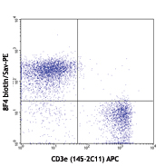
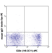
C57BL/6 splenocytes stained with biotin anti-mouse CD272 (clone 8F4)(left) or biotin anti-mouse IgG1 isotype control (right), followed by PE Streptavidin and APC anti-mouse CD3e (clone 145-2C11).
Biotin anti-mouse CD56 + Brilliant Violet 421™ Streptavidin
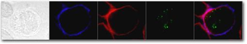
Human NK cell line NK92 fixed and stained with biotin anti-CD56 (clone HCD56) and Brilliant Violet 421™Streptavidin (blue), phalloidin Alexa Fluor® 568 (red), and anti-perforin FITC (green). The first image is the DIC view (far left). The final image (far right) shows the merged fluorescence of the three stains. Data provided by Emily Mace and Jordan Orange, Children's Hospital of Philadelphia.
HLA-A2/peptide Tetramer + PE Streptavidin or Brilliant Violet 421™ Streptavidin
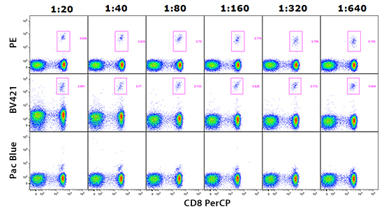
Human PBMCs were stained with PerCP anti-human CD8 and labeled with NLVPMVATV-loaded human HLA-A2 tetramer bound to PE Streptavidin, Brilliant Violet 421™ Streptavidin, or Pacific Blue™ Streptavidin. The data shows positive staining with a wide range of tetramer dilutions for PE and BV421™. Data provided by Rick Willis and John Altman, Emory/Yerkes.
Apotracker™ Tetra + Alexa Fluor® 647 Streptavidin
![]()
Heat-induced 50% dead C57BL/6 splenocytes (left) or untreated splenocytes (right) stained with Helix NP™ Blue and Apotracker™ Tetra Alexa Fluor® 647.
ProductsHere


Follow Us