 Login/Register


- Clone
- 4B10 (See other available formats)
- Regulatory Status
- RUO
- Other Names
- T-box expressed in T cells, T box 21, TBLYM
- Isotype
- Mouse IgG1, κ
- Ave. Rating
- Submit a Review
- Product Citations
- publications

-

Total cell lysate from PBMC (lane 1, 15 µg) and PBMC treated with 5 µg/mL CD3 and 2 µg/mL CD28 (lane 2, 15 µg) were resolved by electrophoresis (4-12% Bis-Tris), transferred to nitrocellulose, and probed with purified anti-T-bet antibody (clone 4B10). Proteins were visualized using an HRP Goat anti-mouse IgG Antibody and chemiluminescence detection. Direct-Blot™ HRP anti-β-actin antibody (clone 2F1-1) was used as a loading control. -

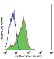
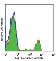
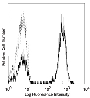
Human peripheral blood lymphocytes were surface stained with CD3 (clone OKT3) APC and then treated with True-Nuclear™ Transcription Factor Buffer Set. Cells were then stained with purified T-bet (clone 4B10) (left) or purified mouse IgG1, κ isotype control (right), followed by anti-mouse IgG1 PE. -

Total lysates (15µg protein) from Daudi, K562 and NK-92 cells were resolved by electrophoresis (4-20% Tris-glycine gel), transferred to nitrocellulose, and probed with 1:500 diluted (1µg/mL) purified anti- T-bet antibody (4B10, upper). Proteins were visualized using an HRP goat anti-mouse-IgG secondary antibody and chemiluminescence detection. Direct-Blot™ HRP anti-β-actin Antibody was used as a loading control (lower). Lane M: Molecular weight ladder.
| Cat # | Size | Price | Quantity Check Availability | Save | ||
|---|---|---|---|---|---|---|
| 644801 | 25 µg | $101 | ||||
| 644802 | 100 µg | $221 | ||||
T-bet, also known as T-box transcription factor T-bet, is considered to be a "master regulator" of Th1 lymphoid development controlling the production of the cytokine IFN-γ. T-bet is widely expressed in hematopoietic cells including stem cells, NK cells, B cells, and T cells. T-bet is critical for the control of microbial pathogens, and knockout animals show multiple physiologic and inflammatory features characteristic of asthma. T-bet expression is optimally observed after IL-12 stimulation and can be suppressed by addition of the Th2 cytokine IL-4 or neutralization of IL-12.
Product DetailsProduct Details
- Verified Reactivity
- Human, Mouse
- Antibody Type
- Monoclonal
- Host Species
- Mouse
- Formulation
- Phosphate-buffered solution, pH 7.2, containing 0.09% sodium azide.
- Preparation
- The antibody was purified by affinity chromatography.
- Concentration
- 0.5 mg/ml
- Storage & Handling
- The antibody solution should be stored undiluted between 2°C and 8°C.
- Application
-
WB, ICFC - Quality tested
IHC-F, IP - Reported in the literature, not verified in house - Recommended Usage
-
Each lot of this antibody is quality control tested by Western blotting and intracelluar immunofluorescent staining using our True-Nuclear™ Transcription Factor Staining Protocol. For Western blotting, the suggested use is 1.0 to 2.0 µg per ml. For flow cytometric staining, the suggested use of this reagent is 1.0 µg per million cells in a staining volume of 100 µl. It is recommended that the reagent be titrated for optimal performance for each application.
- Application Notes
-
Additional reported applications (for the relevant formats) include: immunoprecipitation2 and immunofluorescence microscopy3.
NOTE: For flow cytometric staining with this clone, True-Nuclear™ Transcription Factor Buffer Set (Cat. No. 424401) offers improved staining and is highly recommended over the Foxp3 Fix/Perm Buffer Set (Cat. No. 421403) and the Nuclear Factor Fixation and Permeabilization Buffer Set (Cat. No. 422601). -
Application References
(PubMed link indicates BioLegend citation) -
- Szabo SJ, et al. 2000. Cell 100:655. (ICFC, WB)
- Hwang ES, et al. 2005. J. Exp. Med. 202:1289. (ICFC, WB, IP)
- Neurath MF, et al. 2002. J. Exp. Med. 195:1129. (IF)
- Hsieh CY, et al. 2012. J Pharmacol Exp. 343:125. PubMed.
- Product Citations
-
- RRID
-
AB_1595608 (BioLegend Cat. No. 644801)
AB_1595503 (BioLegend Cat. No. 644802)
Antigen Details
- Structure
- T-box transcription factor, approximately 58 kD.
- Distribution
-
Nuclear; expressed in T cells, hematopoietic stem cells, NK cells, B cells, lung, spleen.
- Function
- Th1-specific T-box transcription factor controlling expression of the hallmark Th1 cytokine, interferon gamma (IFN-γ). T-bet expression is critical for the control of microbial pathogens.
- Cell Type
- B cells, Hematopoietic stem and progenitors, NK cells, T cells, Tregs
- Biology Area
- Cell Biology, Immunology, Transcription Factors
- Molecular Family
- Nuclear Markers
- Antigen References
-
1. Szabo SJ, et al. 2000. Cell 100:655.
2. Szabo SJ, et al. 2002. Science 295:338.
3. Finotto S, et al. 2002. Science 295:336.
4. Mullen AC, et al. 2001. Science 292:1907. - Gene ID
- 30009 View all products for this Gene ID
- UniProt
- View information about T-bet on UniProt.org

Related FAQs
Other Formats
View All T-bet Reagents Request Custom Conjugation| Description | Clone | Applications |
|---|---|---|
| APC anti-T-bet | 4B10 | ICFC |
| Purified anti-T-bet | 4B10 | WB,ICFC,IHC-F,IP |
| Alexa Fluor® 647 anti-T-bet | 4B10 | ICFC |
| PerCP/Cyanine5.5 anti-T-bet | 4B10 | ICFC |
| Pacific Blue™ anti-T-bet | 4B10 | ICFC |
| PE anti-T-bet | 4B10 | ICFC |
| Brilliant Violet 711™ anti-T-bet | 4B10 | ICFC |
| FITC anti-T-bet | 4B10 | ICFC |
| Brilliant Violet 421™ anti-T-bet | 4B10 | ICFC |
| Brilliant Violet 605™ anti-T-bet | 4B10 | ICFC |
| PE/Cyanine7 anti-T-bet | 4B10 | ICFC |
| PE/Dazzle™ 594 anti-T-bet | 4B10 | ICFC |
| Purified anti-T-bet (Maxpar® Ready) | 4B10 | WB,CyTOF® |
| Alexa Fluor® 488 anti-T-bet | 4B10 | ICFC |
| Direct-Blot™ HRP anti-T-bet | 4B10 | WB |
| Brilliant Violet 785™ anti-T-bet | 4B10 | ICFC |
| Alexa Fluor® 594 anti-T-bet | 4B10 | ICFC,ICC |
| KIRAVIA Blue 520™ anti-T-bet | 4B10 | ICFC |
| PE/Fire™ 810 anti-T-bet | 4B10 | ICFC |
Customers Also Purchased
Compare Data Across All Formats
This data display is provided for general comparisons between formats.
Your actual data may vary due to variations in samples, target cells, instruments and their settings, staining conditions, and other factors.
If you need assistance with selecting the best format contact our expert technical support team.
-
APC anti-T-bet

Human peripheral blood lymphocytes were surface stained with... 
-
Purified anti-T-bet

Total cell lysate from PBMC (lane 1, 15 µg) and PBMC treated... 
Human peripheral blood lymphocytes were surface stained with... 
Total lysates (15µg protein) from Daudi, K562 and NK-92 cell... -
Alexa Fluor® 647 anti-T-bet

Human peripheral blood lymphocytes were surface stained with... 
-
PerCP/Cyanine5.5 anti-T-bet

Human peripheral blood lymphocytes were surface stained with... -
Pacific Blue™ anti-T-bet

Human peripheral blood lymphocytes were surface stained with... 
-
PE anti-T-bet

Human peripheral blood lymphocytes were surface stained with... 
-
Brilliant Violet 711™ anti-T-bet

Human peripheral blood lymphocytes were surface stained with... 
-
FITC anti-T-bet

Human peripheral blood lymphocytes were surface stained with... 
-
Brilliant Violet 421™ anti-T-bet

Human peripheral blood lymphocytes were surface stained with... 
-
Brilliant Violet 605™ anti-T-bet

Human peripheral blood lymphocytes were surface stained with... 
-
PE/Cyanine7 anti-T-bet

Human peripheral blood lymphocytes were surface stained with... -
PE/Dazzle™ 594 anti-T-bet

Human peripheral blood lymphocytes were surface stained with... 
-
Purified anti-T-bet (Maxpar® Ready)

Human PBMCs were fixed, permeabilized, and stained with 154S... -
Alexa Fluor® 488 anti-T-bet

Human peripheral blood lymphocytes were surface stained with... 
-
Direct-Blot™ HRP anti-T-bet

Total cell lysate from PBMC (lane 1, 15 µg) and PBMC treated... -
Brilliant Violet 785™ anti-T-bet

Human peripheral blood lymphocytes were stained with CD3 APC... 
-
Alexa Fluor® 594 anti-T-bet

NK-92 cells were fixed with 4% paraformaldehyde (PFA) for 15... 
NK-92 cells were fixed with 4% paraformaldehyde (PFA) for 15... 
PBMCs were surface stained with Alexa Fluor® 488 anti-CD3 (C... -
KIRAVIA Blue 520™ anti-T-bet

Human peripheral blood lymphocytes were surface stained with... -
PE/Fire™ 810 anti-T-bet

Human peripheral blood lymphocytes were surface stained with...


Follow Us