 Login/Register


- Clone
- LCK-01 (See other available formats)
- Regulatory Status
- RUO
- Other Names
- Proto-oncogene tyrosine kinase lck, p56-Lck, T-cell specific protein tyrosine kinase
- Isotype
- Mouse IgG2a, κ
- Ave. Rating
- Submit a Review
- Product Citations
- publications

-

Total cell lysates (15 µg total protein) from Jurkat, MOLT4, EL4 cells were resolved by 4-12% Bis-Tris gel electrophoresis, transferred to a PVDF membrane, and probed with 1.0 µg/mL (1:500 dilution) of Purified anti-LCK Antibody, clone LCK-01, overnight at 4°C. Proteins were visualized by chemiluminescence detection using HRP goat anti-mouse IgG Antibody (Cat. No. 405306) at a 1:3000 dilution. Direct-Blot™ HRP anti-GAPDH Antibody (Cat. No. 607904) was used as a loading control at a 1:50000 dilution (lower). Lane M: Molecular Weight marker. -

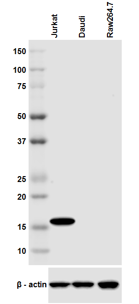
Total cell lysate from Jurkat cells (lane 1, 15 µg), mouse thymus (lane 2, 15 µg) and HeLa cells (lane 3, 15 µg) were resolved by electrophoresis (4-20% Tris-Glycine gel), transferred to nitrocellulose, and probed with purified anti-Lck antibody (clone LCK-01). Proteins were visualized using an HRP Goat anti-mouse IgG Antibody and chemiluminescence detection. Direct-Blot™ HRP anti-β-actin antibody (clone 2F1-1) was used as a loading control. -

Jurkat cells were fixed with 4% PFA for ten minutes, permeabilized with 0.5% Triton X-100 for five minutes, and blocked with 5% FBS for 30 minutes. Then the cells were stained with 2 µg/mL anti-Lck Antibody (clone LCK-01) followed by Alexa Fluor® 594 (red) conjugated goat anti-mouse IgG in blocking buffer for two hours at room temperature. Nuclei were counterstained with DAPI (blue). The image was captured with a 60X objective. -

Whole cell extracts (300 µg total protein) prepared from Jurkat cells were immunoprecipitated overnight with 2.5 µg of Purified mouse IgG1, κ Isotype Control Antibody (Cat. No. 401401) or Purified anti-Lck Antibody, clone LCK-01. The resulting IP fractions and whole cell extract input (5%) were resolved by 4-12% Bis-Tris gel electrophoresis, transferred to a PVDF membrane, and probed with a 1.0 µg/mL (1:500 dilution) of LCK-01. Lane M: Molecular Weight marker.
| Cat # | Size | Price | Quantity Check Availability | Save | ||
|---|---|---|---|---|---|---|
| 628301 | 25 µg | $106 | ||||
| 628302 | 100 µg | $229 | ||||
Lck is a 56 kD tyrosine protein kinase and a member of the src subfamily that contains both an SH2 and SH3 domain. Alternatively spliced isoforms of Lck have been reported. Lck is a cytoplasmic protein bound to CD4 and CD8 in T lymphocytes that participates in antigen-induced T cell activation through phosphorylation of the T cell receptor zeta chain. Lck is activated upon T cell receptor engagement and is involved in the onset of cell cycle progression induced by interleukin-2. Phosphorylation by Csk downregulates the activity of Lck. In addition to CD4 and CD8, Lck has been reported to interact with PI3K through its SH3 domain and tyrosine phosphorylated KHDRBS1/p70 through its SH2 domain. In addition, Lck has been shown to associate with the SAP transmembrane tyrosine phosphatase. The Lck-01 monoclonal antibody recognizes human Lck and has been shown to be useful for Western blotting, immunoprecipitation, immunocytochemistry, and flow cytometry.
Product DetailsProduct Details
- Verified Reactivity
- Human
- Antibody Type
- Monoclonal
- Host Species
- Mouse
- Immunogen
- Peptide corresponding to a.a. 22-36 in human Lck
- Formulation
- This antibody is provided in phosphate-buffered solution, pH 7.2, containing 0.09% sodium azide at 0.5 mg/ml.
- Preparation
- The antibody was purified by affinity chromatography.
- Concentration
- 0.5 mg/ml
- Storage & Handling
- The antibody solution should be stored undiluted between 2°C and 8°C.
- Application
-
WB - Quality tested
ICC, IP - Verified
FC - Reported in the literature, not verified in house - Recommended Usage
-
Each lot of this antibody is quality control tested by Western blotting. Western blotting, suggested working dilution(s): Use 5 µg per 5 ml antibody dilution buffer for each mini-gel. For immunocytochemistry, a concentration range of 1.0 - 5.0 µg/ml is recommended. For immunoprecipitation, the suggested use of this reagent is 2.5 µg per ml. It is recommended that the reagent be titrated for optimal performance for each application.
- Application Notes
-
Does not cross-react with mouse.
Clone may cross-react with other Src tyrosine kinase family members. -
Application References
(PubMed link indicates BioLegend citation) -
- Romagnoli P, et al. 2001. Int. Immunol. 13:305.
- Product Citations
-
- RRID
-
AB_2136337 (BioLegend Cat. No. 628301)
AB_2136336 (BioLegend Cat. No. 628302)
Antigen Details
- Structure
- Belongs to the tyrosine family of kinases, src subfamily. Contains one SH2 domain, 1 SH3 domain. Approximately 56 kD, alternatively spliced isoforms have been reported
- Distribution
-
Cytoplasmic protein, bound to cytoplasmic CD4 and CD8 in T lymphocytes
- Function
- Participates in antigen-induced T cell activation, phosphorylates zeta chain of T cell receptor. Involved in interleukin-2 induced onset of cell cycle progression
- Interaction
- Associates with CD4 and CD8 co-receptors. Binds to PI3K through SH3 interactions, binds to tyrosine phosphorylated KHDRBS1/p70 through SH2 domain. Interacts with SAP transmembrane tyrosine phosphatase
- Modification
- Phosphorylation
- Cell Type
- T cells
- Biology Area
- Cell Biology, Signal Transduction
- Molecular Family
- Protein Kinases/Phosphatase
- Antigen References
-
1. Koga Y, et al. 1986. Eur. J. Immunol. 16:1643.
2. Vogel LB, et al. 1995. Biochem. Biophys. Acta 1264:168.
3. Vogel LB, et al. 1993. Mol. Cell. Biol. 13:7408.
4. Bergaman M, et al. 1992. EMBO J. 11:2919. - Regulation
- Activated upon T cell receptor engagement. Tyrosine phosphorylation by p50 Csk downregulates catalytic activity
- Gene ID
- 3932 View all products for this Gene ID
- UniProt
- View information about Lck on UniProt.org

Related FAQs
Other Formats
View All Lck Reagents Request Custom Conjugation| Description | Clone | Applications |
|---|---|---|
| Purified anti-Lck | LCK-01 | WB,ICC,IP,FC |
| Alexa Fluor® 647 anti-Lck | LCK-01 | ICFC,FC |
Customers Also Purchased

Compare Data Across All Formats
This data display is provided for general comparisons between formats.
Your actual data may vary due to variations in samples, target cells, instruments and their settings, staining conditions, and other factors.
If you need assistance with selecting the best format contact our expert technical support team.
-
Purified anti-Lck

Total cell lysate from Jurkat cells (lane 1, 15 µg), mouse t... 
Jurkat cells were fixed with 4% PFA for ten minutes, permeab... 
Total cell lysates (15 µg total protein) from Jurkat, MOLT4,... 
Whole cell extracts (300 µg total protein) prepared from Jur... -
Alexa Fluor® 647 anti-Lck

Human peripheral blood lymphocytes were surface stained with... 


Follow Us