 Login/Register


- Clone
- A15143K (See other available formats)
- Regulatory Status
- RUO
- Other Names
- Microtubule-associated proteins 1A/1B light chain 3B, MAP1A/MAP1B LC3 B, MAP1A/1B light chain 3 B, MAP1 light chain 3-like protein 2, autophagy-related ubiquitin-like modifier LC3 B
- Isotype
- Mouse IgG2b, κ
- Ave. Rating
- Submit a Review
- Product Citations
- publications

-

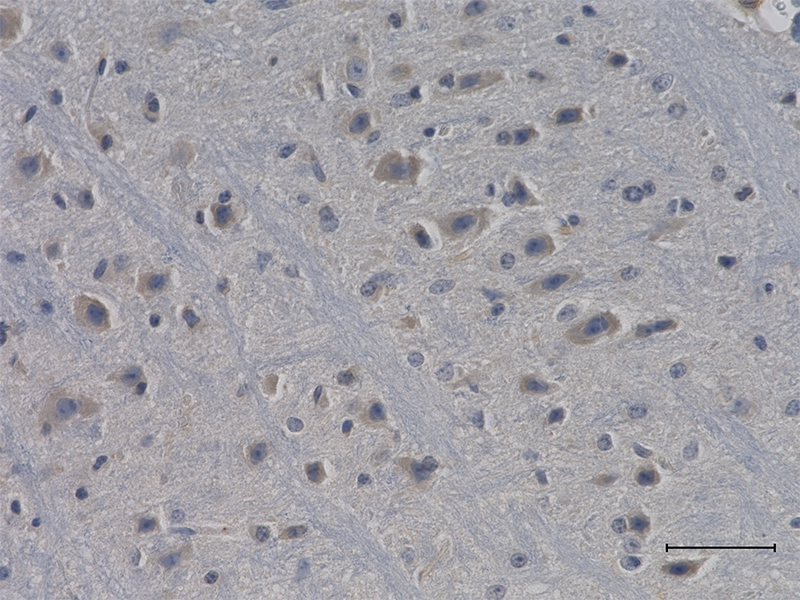
IHC staining of purified anti-LC3 antibody (clone A15143K) on formalin-fixed paraffin-embedded human and rat brain tissues. Following antigen retrieval using Sodium Citrate H.I.E.R., the tissues were incubated with the primary antibody at 5 µg/mL overnight at 4°C. BioLegend’s Ultra-Streptavidin (USA) HRP Detection Kit (Multi-Species, DAB, Cat. No. 929901) was used for detection followed by hematoxylin counterstaining, according to the protocol provided. -

ICC staining of purified anti-LC3 antibody (clone A15143K) on SH-SY5Y neuroblastoma cells. The cells were fixed with 4% PFA, permeabilized with a buffer containing 0.1% Triton X-100 and 0.25% BSA, and blocked with 2% normal goat serum and 0.02% BSA. The cells were then incubated with 1 µg/mL of the primary antibody for three hours at room temperature, followed by 1 µg /mL of Alexa Fluor® 488 goat anti-mouse IgG antibody (Cat. No. 405319) (green). Cells were then incubated with 1 µg/mL of Alexa Fluor® 647 anti-Tubulin β 3 (TUBB3) antibody (Cat. No. 801210) (pink) overnight at 4°C. Nuclei were stained with Hoechst 33342 (blue) at 5 µg/mL for 10 minutes at room temperature. Images were captured with a 60X objective.
| Cat # | Size | Price | Quantity Check Availability | Save | ||
|---|---|---|---|---|---|---|
| 848801 | 25 µg | $112 | ||||
| 848802 | 100 µg | $276 | ||||
Microtubule-associated protein light chain 3, commonly known as LC3, is a central protein in the autophagy pathway and functions in autophagosome biogenesis. LC3 is a member of the ATG8 protein family and expressed as three isoforms: LC3A (MAP1LC3A), LC3B (MAP1LC3B), and LC3C (MAP1LC3C). Newly synthesized LC3 is hydrolyzed by the cysteine protease ATG4B to produce the active cytosolic form termed LC3I. Through a series of reactions involving other ATGs, LC3I becomes attached to phosphatidylethanolamine. This lipid modified form is termed LC3II, and is involved in autophagosomes membrane expansion and fusion events. LC3 is widely used as a marker for autophagosomes. Defects in autophagy machinery have been associated with multiples diseases including neurodegenerative disorders.
Product DetailsProduct Details
- Verified Reactivity
- Human, Mouse
- Antibody Type
- Monoclonal
- Host Species
- Mouse
- Immunogen
- A synthetic peptide corresponding to N-terminal region of human LC3B protein (between amino acid residues 1-50) conjugated to KLH.
- Formulation
- Phosphate-buffered solution, pH 7.2, containing 0.09% sodium azide.
- Preparation
- The antibody was purified by affinity chromatography.
- Concentration
- 0.5 mg/mL
- Storage & Handling
- The antibody solution should be stored undiluted between 2°C and 8°C.
- Application
-
IHC-P - Quality tested
ICC - Verified
WB - Reported in the literature, not verified in house - Recommended Usage
-
Each lot of this antibody is quality control tested by formalin-fixed paraffin-embedded immunohistochemical staining. For immunohistochemistry, a concentration range of 1.0 - 5.0 µg/mL is suggested. For immunocytochemistry, a concentration range of 0.5 - 1.0 µg/mL is suggested It is recommended that the reagent be titrated for optimal performance for each application.
- Application Notes
-
This antibody cross-reacts with LC3A and LC3B isoforms.
Additional reported applications (for the relevant formats) include: western blotting - Product Citations
-
- RRID
-
AB_2650713 (BioLegend Cat. No. 848801)
AB_2650714 (BioLegend Cat. No. 848802)
Antigen Details
- Structure
- LC3 is a 125 amino acid protein with a molecular mass of 14.7 kD.
- Distribution
-
Tissue distribution: brain, heart, skeletal muscle, blood and testis.
Cellular distribution: cytosol, mitochondria, cytoskeleton, endoplasmic reticulum, cytoplasmic vesicles, autophagosome. - Function
- LC3 proteins play a key role in the selective recruitment of autophagic cargoes into autophagosomes, and serve as docking sites for adaptor proteins.
- Biology Area
- Cell Biology, Neurodegeneration, Neuroscience, Neuroscience Cell Markers, Protein Trafficking and Clearance
- Molecular Family
- Autophagosome Markers
- Antigen References
-
1. Lee J, et al. 2012. Biochem. J. 441(2):523. PubMed
2. Martini-Stoica H, et al. 2016. Trends Neurosci. 39(4)221. PubMed - Gene ID
- 81631 View all products for this Gene ID
- UniProt
- View information about LC3 on UniProt.org

Related FAQs
Other Formats
View All LC3 Reagents Request Custom Conjugation| Description | Clone | Applications |
|---|---|---|
| Purified anti-LC3 | A15143K | IHC-P,ICC |
Customers Also Purchased
Compare Data Across All Formats
This data display is provided for general comparisons between formats.
Your actual data may vary due to variations in samples, target cells, instruments and their settings, staining conditions, and other factors.
If you need assistance with selecting the best format contact our expert technical support team.


Follow Us