 Login/Register


- Clone
- W17211A (See other available formats)
- Regulatory Status
- RUO
- Other Names
- MKI67, Proliferation Marker Protein Ki-67, Antigen Ki67
- Isotype
- Rat IgG2a, κ
- Ave. Rating
- Submit a Review
- Product Citations
- publications

-

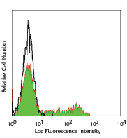
Human paraffin-embedded intestine tissue slices were prepared with a standard protocol of deparaffinization and rehydration. Antigen retrieval was done with Citrate-Buffered 1X pH 6.0 at 95°C for 40 minutes. Tissue was washed with PBS/0.05% Tween 20 twice for five minutes, permerilized with 0.5% Triton X-100 for 10 minutes and blocked with 5% FBS and 0.2% gelatin for 30 minutes. Then, the tissue was stained with 10 µg/mL of purified anti-human Ki-67 (clone W17211A) antibody overnight at 4°C. On the next day, tissue was incubate with Alexa Fluor® 594 goat anti-rat IgG (clone poly4054) antibody (red). Nuclei were counterstained with DAPI (blue). The image was scanned with a 10X objective and stitched with MetaMorph® software. -

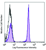
Human paraffin-embedded tonsil tissue slices were prepared with a standard protocol of deparaffinization and rehydration. Antigen retrieval was done with Citrate-Buffered 1X pH 6.0 at 95°C for 40 minutes. Tissue was washed with PBS/0.05% Tween 20 twice for five minutes, permerilized with 0.5% Triton X-100 for 10 minutes and blocked with 5% FBS and 0.2% gelatin for 30 minutes. Then, the tissue was stained with 10 µg/mL of purified anti-human Ki-67 (clone W17211A) antibody overnight at 4°C. On the next day, tissue was incubate with Alexa Fluor® 594 goat anti-rat IgG (clone poly4054) antibody (red). Nuclei were counterstained with DAPI (blue). The image was scanned with a 10X objective and stitched with MetaMorph® software.
| Cat # | Size | Price | Quantity Check Availability | Save | ||
|---|---|---|---|---|---|---|
| 398502 | 100 µg | $136 | ||||
Antigen Ki-67 is a nuclear protein expressed as two isoforms with molecular weights of 395 and 345 kD. Both isoforms contain one forkhead-associated domain and 16 concatenated "Ki-67 repeats," each containing the epitope recognized by the mAb Ki-67. The antigen Ki-67 interacts with Hklp2, hNIFK, and chromobox protein homolog 1, 3, and 5. Ki-67 is required for cell proliferation and its expression is restricted to the phases G1, S, G2, and M of the cell cycle. This characteristic makes Ki-67 an excellent marker for proliferating cells and is commonly used as one of the prognostic factors in cancer studies. Ki-67 has also been used to study myocyte proliferation after myocardial infarction as well as lymphocyte proliferation during infection, and has been used in neurons of patients with different neuropathologies.
Product DetailsProduct Details
- Verified Reactivity
- Human
- Antibody Type
- Monoclonal
- Host Species
- Rat
- Immunogen
- Human Ki-67 recombinant protein
- Formulation
- Phosphate-buffered solution, pH 7.2, containing 0.09% sodium azide
- Preparation
- The antibody was purified by affinity chromatography.
- Concentration
- 0.5 mg/mL
- Storage & Handling
- The antibody solution should be stored undiluted between 2°C and 8°C.
- Application
-
IHC-P - Quality tested
- Recommended Usage
-
Each lot of this antibody is quality control tested by formalin-fixed paraffin-embedded immunohistochemical staining. For immunohistochemistry, a concentration range of 5 - 10 µg/mL is suggested. It is recommended that the reagent be titrated for optimal performance for each application.
- RRID
-
AB_2820065 (BioLegend Cat. No. 398502)
Antigen Details
- Structure
- Two isoforms with molecular weights of 395 and 345 kD, one forkhead-associated domain, 16 concatenated Ki-67 repeats, located in nucleus
- Distribution
-
Expressed in the phases G1, S, G2, and M of the cell cycle
- Function
- Required for cell proliferation
- Interaction
- Chromobox protein homolog 1, 3 and 5, Hklp2, and hNIFK
- Biology Area
- Cell Biology, Cell Cycle/DNA Replication, DNA Repair/Replication
- Molecular Family
- Nuclear Markers
- Antigen References
-
- Byeon IJ, et al. 2005. Nat. Struct. Mol. Biol. 12:987.
- Yerushalmi R, et al. 2010. Lancet. Oncol. 11:174.
- Drazen JM. et al. 2001. N. Engl. J. Med. 344:1750.
- Sachsenberg N, et al. 1998. J. Exp. Med. 187:1295.
- Nagy Z, et al. 1997. Acta. Neuropathol. 93:294.
- Gene ID
- 4288 View all products for this Gene ID
- UniProt
- View information about Ki-67 on UniProt.org

Related FAQs
Other Formats
View All Ki-67 Reagents Request Custom Conjugation| Description | Clone | Applications |
|---|---|---|
| Purified anti-human Ki-67 | W17211A | IHC-P |
| TotalSeq™-Bn1331 anti-human Ki-67 | W17211A | SB |
Customers Also Purchased
Compare Data Across All Formats
This data display is provided for general comparisons between formats.
Your actual data may vary due to variations in samples, target cells, instruments and their settings, staining conditions, and other factors.
If you need assistance with selecting the best format contact our expert technical support team.
-
Purified anti-human Ki-67

Human paraffin-embedded intestine tissue slices were prepare... 
Human paraffin-embedded tonsil tissue slices were prepared w... -
TotalSeq™-Bn1331 anti-human Ki-67


Follow Us