 Login/Register


- Clone
- P1H12 (See other available formats)
- Regulatory Status
- RUO
- Workshop
- HCDM listed
- Other Names
- Muc-18, MCAM, Mel-CAM, S-endo
- Isotype
- Mouse IgG1, κ
- Ave. Rating
- Submit a Review
- Product Citations
- publications

-

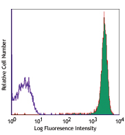
Human cervical cancer cell line, HeLa was stained with CD146 (clone P1H12) PE/Cyanine7 (filled histogram) or mouse IgG1, κ PE/Cyanine7 isotype control (open histogram).
| Cat # | Size | Price | Quantity Check Availability | Save | ||
|---|---|---|---|---|---|---|
| 361007 | 25 tests | $159 | ||||
| 361008 | 100 tests | $370 | ||||
CD146 is a 118 kD integral transmembrane glycoprotein that is also known as MUC18, S-Endo, MCAM, and Mel-CAM (melanoma cell adhesion molecule). It belongs to the immunoglobulin superfamily. CD146 is expressed on melanoma cells, epithelial cells, endothelial cells, fibroblasts, activated T cells, multipotent mesenchymal stromal cells, and activated keratinocytes. CD146 mediates heterophilic cell adhesion and regulates monocyte transendothelial migration. The ligand of CD146 remains to be identified.
Product DetailsProduct Details
- Verified Reactivity
- Human
- Reported Reactivity
- Mouse, Dog, Rabbit
- Antibody Type
- Monoclonal
- Host Species
- Mouse
- Immunogen
- Cultured human umbilical cells
- Formulation
- Phosphate-buffered solution, pH 7.2, containing 0.09% sodium azide and BSA (origin USA)
- Preparation
- The antibody was purified by affinity chromatography and conjugated with PE/Cyanine7 under optimal conditions.
- Concentration
- Lot-specific (to obtain lot-specific concentration and expiration, please enter the lot number in our Certificate of Analysis online tool.)
- Storage & Handling
- The antibody solution should be stored undiluted between 2°C and 8°C, and protected from prolonged exposure to light. Do not freeze.
- Application
-
FC - Quality tested
- Recommended Usage
-
Each lot of this antibody is quality control tested by immunofluorescent staining with flow cytometric analysis. For flow cytometric staining, the suggested use of this reagent is 5 µl per million cells in 100 µl staining volume or 5 µl per 100 µl of whole blood.
- Excitation Laser
-
Blue Laser (488 nm)
Green Laser (532 nm)/Yellow-Green Laser (561 nm)
- Application Notes
-
Additional reported applications (for the relevant formats of this clone) include: Western blotting3 and IHC1,5.
Clone P1H12 cross-reacts to Dog2, 5. - Additional Product Notes
-
BioLegend is in the process of converting the name PE/Cy7 to PE/Cyanine7. The dye molecule remains the same, so you should expect the same quality and performance from our PE/Cyanine7 products. Please contact Technical Service if you have any questions.
-
Application References
(PubMed link indicates BioLegend citation) -
- Solovey A, et al. 1997. N. Engl. J. Med. 337:1584. (FC, IHC)
- Lamerato-Kozicki AR, et al. 2006. Exp. Hematol. 34:870. (FC)
- Balint K, et al. 2005. J. Clin. Invest. 115:3166. (WB)
- Neskey DM, et al. 2008. J. Exp. Clin. Cancer Res. 27:61. (ELISA)
- Kamstock D, et al. 2006. Cancer Gene Therap. 13:306. (IHC)
- Product Citations
-
- RRID
-
AB_2562982 (BioLegend Cat. No. 361007)
AB_2562983 (BioLegend Cat. No. 361008)
Antigen Details
- Structure
- 118 kD integral glycoprotein, Ig superfamily
- Distribution
-
Melanoma cells, epithelial cells, endothelial cells, activated T cells, multipotent mesenchymal stromal cells
- Function
- Adhesion, monocytes transendothelial migration
- Cell Type
- Endothelial cells, Epithelial cells, Mesenchymal cells, Mesenchymal Stem Cells, T cells
- Biology Area
- Cell Biology, Immunology, Innate Immunity, Neuroscience, Neuroscience Cell Markers, Stem Cells
- Molecular Family
- Adhesion Molecules, CD Molecules
- Antigen References
-
1. Pickl WF, et al. 1997. J. Immunol. 158:2107.
2. Weninger W, et al. 2000. J. Invest. Dermatol. 115:219.
3. Sorrentino A, et al. 2008. Exp. Hematol. 36:1035.
4. Bardin N, et al. 2009. Arterioscler. Thromb. Vasc. Biol. 29:746. - Gene ID
- 4162 View all products for this Gene ID
- UniProt
- View information about CD146 on UniProt.org
Related Pages & Pathways
Pathways


Related FAQs
Other Formats
View All CD146 Reagents Request Custom Conjugation| Description | Clone | Applications |
|---|---|---|
| PE anti-human CD146 | P1H12 | FC |
| Purified anti-human CD146 | P1H12 | FC,IHC-F,WB |
| Brilliant Violet 421™ anti-human CD146 | P1H12 | FC |
| PE/Cyanine7 anti-human CD146 | P1H12 | FC |
| PerCP/Cyanine5.5 anti-human CD146 | P1H12 | FC |
| FITC anti-human CD146 | P1H12 | FC |
| Alexa Fluor® 647 anti-human CD146 | P1H12 | FC |
| APC anti-human CD146 | P1H12 | FC |
| TotalSeq™-A0134 anti-human CD146 | P1H12 | PG |
| Brilliant Violet 605™ anti-human CD146 | P1H12 | FC |
| Brilliant Violet 510™ anti-human CD146 | P1H12 | FC |
| Alexa Fluor® 488 anti-human CD146 | P1H12 | FC |
| APC/Fire™ 750 anti-human CD146 | P1H12 | FC |
| PE/Dazzle™ 594 anti-human CD146 | P1H12 | FC |
| Brilliant Violet 785™ anti-human CD146 | P1H12 | FC |
| Brilliant Violet 711™ anti-human CD146 | P1H12 | FC |
| PE/Cyanine5 anti-human CD146 | P1H12 | FC |
| Biotin anti-human CD146 | P1H12 | FC |
| TotalSeq™-C0134 anti-human CD146 | P1H12 | PG |
| TotalSeq™-B0134 anti-human CD146 Antibody | P1H12 | PG |
| APC/Cyanine7 anti-human CD146 | P1H12 | FC |
Customers Also Purchased
Compare Data Across All Formats
This data display is provided for general comparisons between formats.
Your actual data may vary due to variations in samples, target cells, instruments and their settings, staining conditions, and other factors.
If you need assistance with selecting the best format contact our expert technical support team.
-
PE anti-human CD146

Human cervical cancer cell line HeLa was stained with CD146 ... -
Purified anti-human CD146

Human cervical cancer cell line HeLa was stained with purifi... -
Brilliant Violet 421™ anti-human CD146

Human cervical cancer cell line HeLa was stained with CD146 ... -
PE/Cyanine7 anti-human CD146

Human cervical cancer cell line, HeLa was stained with CD146... -
PerCP/Cyanine5.5 anti-human CD146

Human cervical cancer cells (HeLa cell line) were stained wi... -
FITC anti-human CD146

Human cervical cancer cell line, HeLa, was stained with anti... -
Alexa Fluor® 647 anti-human CD146

Human cervical cancer cell line, HeLa, was stained with anti... -
APC anti-human CD146

Human cervical cancer cell line, HeLa, was stained with CD14... -
TotalSeq™-A0134 anti-human CD146
-
Brilliant Violet 605™ anti-human CD146

Human cervical cancer cell line, HeLa, was stained with CD14... -
Brilliant Violet 510™ anti-human CD146

Human cervical cancer cell line, HeLa, was stained with CD14... -
Alexa Fluor® 488 anti-human CD146

Human cervical cancer cell line, HeLa, was stained with CD14... -
APC/Fire™ 750 anti-human CD146

Human cervical cancer cell line, HeLa, was stained with CD14... -
PE/Dazzle™ 594 anti-human CD146

Human cervical cancer cell line, HeLa, was stained with CD14... -
Brilliant Violet 785™ anti-human CD146

Human cervical cancer cell line, HeLa, was stained with CD14... -
Brilliant Violet 711™ anti-human CD146

Human cervical cancer cell line, HeLa, was stained with CD14... -
PE/Cyanine5 anti-human CD146

Human cervical cancer cell line, HeLa, was stained with CD14... -
Biotin anti-human CD146

Human cervical cancer cell line, HeLa, was stained with CD14... -
TotalSeq™-C0134 anti-human CD146
-
TotalSeq™-B0134 anti-human CD146 Antibody
-
APC/Cyanine7 anti-human CD146

Human cervical cancer cell line, HeLa, was stained with anti...


Follow Us