 Login/Register


- Clone
- ZET (See other available formats)
- Regulatory Status
- RUO
- Other Names
- GPR5, CCXCR1, mXCR1, Lymphotactin receptor, SCM1 receptor, XC chemokine receptor 1
- Isotype
- Mouse IgG2b, κ
- Ave. Rating
- Submit a Review
- Product Citations
- publications

-

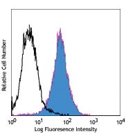
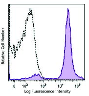
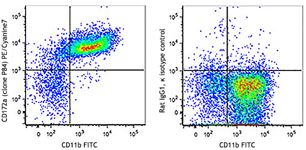
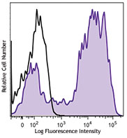
Cells from collagenase-digested C57BL/6 mouse spleen were stained with CD11c Brilliant Violet 421™, CD8α APC and XCR1 (clone ZET) PE (top) or mouse IgG2b, κ PE isotype control (bottom). Data shown was gated on the CD11c+ population. -

| Cat # | Size | Price | Quantity Check Availability | Save | ||
|---|---|---|---|---|---|---|
| 148203 | 25 µg | $153 | ||||
| 148204 | 100 µg | $335 | ||||
XCR1, also known as GPR5 or CCXCR1, is a 38 kD, G-protein coupled, seven transmembrane receptor, and the only member of the "C" chemokine receptor family. XCR1 mediates chemotaxis of XCL1 and XCL2 (lymphotactin-1 and -2), and defines a subset of CD8α+ conventional dendritic cells capable of antigen cross-presentation.
Product DetailsProduct Details
- Verified Reactivity
- Mouse, Rat
- Antibody Type
- Monoclonal
- Host Species
- Mouse
- Formulation
- Phosphate-buffered solution, pH 7.2, containing 0.09% sodium azide.
- Preparation
- The antibody was purified by affinity chromatography and conjugated with PE under optimal conditions.
- Concentration
- 0.2 mg/ml
- Storage & Handling
- The antibody solution should be stored undiluted between 2°C and 8°C, and protected from prolonged exposure to light. Do not freeze.
- Application
-
FC - Quality tested
- Recommended Usage
-
Each lot of this antibody is quality control tested by immunofluorescent staining with flow cytometric analysis. For flow cytometric staining, the suggested use of this reagent is ≤0.25 µg per million cells in 100 µl volume. It is recommended that the reagent be titrated for optimal performance for each application.
- Excitation Laser
-
Blue Laser (488 nm)
Green Laser (532 nm)/Yellow-Green Laser (561 nm)
- Application Notes
-
Additional reported applications (for the relevant formats) include: immunohistochemical staining on frozen tissues2.
-
Application References
(PubMed link indicates BioLegend citation) -
- Gurka S, et al. 2015. Front Immunol. 6:35. (FC)
- Kitazawa Y, et al. 2019. Front Immunol. 10:1195. (FC, IHC-F)
- Product Citations
-
- RRID
-
AB_2563842 (BioLegend Cat. No. 148203)
AB_2563843 (BioLegend Cat. No. 148204)
Antigen Details
- Structure
- Only member of the "C" chemokine receptor family, seven transmembrane receptor, coupled to G protein, 38 kD.
- Distribution
-
Subset of CD8α+ conventional dendritic cells.
- Function
- Mediates chemotaxis to XCL1, defines a population of CD8α+ dendritic cells capable of antigen cross-presentation to CD8+ T cells.
- Ligand/Receptor
- XCL1 and XCL2.
- Cell Type
- Dendritic cells
- Biology Area
- Cell Biology, Immunology, Innate Immunity, Signal Transduction
- Molecular Family
- Cytokine/Chemokine Receptors, GPCR
- Antigen References
-
1. Yamazaki C, et al. 2013. J. Immunol. 190:6071.
2. Shimizu K, et al. 2013. J. Immunol. 190:5609.
3. Bachem A, et al. 2012. Front. Immunol. 3:214.
4. Crozat K, et al. 2011. J. Immunol. 187:4411. - Gene ID
- 23832 View all products for this Gene ID 301086 View all products for this Gene ID
- UniProt
- View information about XCR1 on UniProt.org
Related Pages & Pathways
Pathways


Related FAQs
- What type of PE do you use in your conjugates?
- We use R-PE in our conjugates.
Other Formats
View All XCR1 Reagents Request Custom Conjugation| Description | Clone | Applications |
|---|---|---|
| PE anti-mouse/rat XCR1 | ZET | FC |
| Purified anti-mouse/rat XCR1 | ZET | FC,IHC-F |
| APC anti-mouse/rat XCR1 | ZET | FC |
| PerCP/Cyanine5.5 anti-mouse/rat XCR1 | ZET | FC |
| FITC anti-mouse/rat XCR1 | ZET | FC |
| Biotin anti-mouse/rat XCR1 | ZET | FC |
| Alexa Fluor® 647 anti-mouse/rat XCR1 | ZET | FC |
| Brilliant Violet 421™ anti-mouse/rat XCR1 | ZET | FC |
| Brilliant Violet 510™ anti-mouse/rat XCR1 | ZET | FC |
| Brilliant Violet 650™ anti-mouse/rat XCR1 | ZET | FC |
| APC/Cyanine7 anti-mouse/rat XCR1 | ZET | FC |
| Brilliant Violet 785™ anti-mouse/rat XCR1 | ZET | FC |
| TotalSeq™-A0568 anti-mouse/rat XCR1 | ZET | PG |
| TotalSeq™-C0568 anti-mouse/rat XCR1 | ZET | PG |
| TotalSeq™-B0568 anti-mouse/rat XCR1 Antibody | ZET | PG |
| PE/Dazzle™ 594 anti-mouse/rat XCR1 | ZET | FC |
| Brilliant Violet 605™ anti-mouse/rat XCR1 | ZET | FC |
| Spark UV™ 387 anti-mouse/rat XCR1 | ZET | FC |
Customers Also Purchased
Compare Data Across All Formats
This data display is provided for general comparisons between formats.
Your actual data may vary due to variations in samples, target cells, instruments and their settings, staining conditions, and other factors.
If you need assistance with selecting the best format contact our expert technical support team.
-
PE anti-mouse/rat XCR1

Cells from collagenase-digested C57BL/6 mouse spleen were st... 
-
Purified anti-mouse/rat XCR1

Cells from collagenase-digested C57BL/6 mouse spleen were st... 
-
APC anti-mouse/rat XCR1

Cells from collagenase-digested C57BL/6 mouse spleen were st... 
-
PerCP/Cyanine5.5 anti-mouse/rat XCR1

Mouse spleen cells, from collagenase-digested C57BL/6, were ... -
FITC anti-mouse/rat XCR1

Mouse spleen cells, from collagenase-digested C57BL/6, were ... 
-
Biotin anti-mouse/rat XCR1

Mouse spleen cells, from collagenase-digested C57BL/6, were ... 
-
Alexa Fluor® 647 anti-mouse/rat XCR1

Mouse spleen cells, from collagenase-digested C57BL/6, were ... 
-
Brilliant Violet 421™ anti-mouse/rat XCR1

Cells from collagenase-digested C57BL/6 mouse spleen were st... 
-
Brilliant Violet 510™ anti-mouse/rat XCR1

Cells from collagenase-digested C57BL/6 mouse spleen were st... 
-
Brilliant Violet 650™ anti-mouse/rat XCR1

Cells from collagenase-digested C57BL/6 mouse spleen were st... 
-
APC/Cyanine7 anti-mouse/rat XCR1

Cells from collagenase-digested C57BL/6 mouse spleen were st... -
Brilliant Violet 785™ anti-mouse/rat XCR1

Cells from collagenase-digested C57BL/6 mouse spleen were st... -
TotalSeq™-A0568 anti-mouse/rat XCR1
-
TotalSeq™-C0568 anti-mouse/rat XCR1
-
TotalSeq™-B0568 anti-mouse/rat XCR1 Antibody
-
PE/Dazzle™ 594 anti-mouse/rat XCR1

Cells from collagenase-digested C57BL/6 mouse spleen were st... -
Brilliant Violet 605™ anti-mouse/rat XCR1

Cells from collagenase-digested C57BL/6 mouse spleen were st... -
Spark UV™ 387 anti-mouse/rat XCR1

Cells from collagenase-digested C57BL/6 mouse spleen were st...


Follow Us