 Login/Register


- Clone
- RMT3-23 (See other available formats)
- Regulatory Status
- RUO
- Other Names
- T cell immunoglobulin and mucin domain containing 3 protein, hepatitis virus cellular receptor 2, CD366
- Isotype
- Rat IgG2a, κ
- Ave. Rating
- Submit a Review
- Product Citations
- publications

-

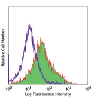
C57BL/6 mouse splenocytes were stained with anti-mouse CD3ε (clone 145-2C11) PerCP/Fire™ 780 and anti-mouse CD366 (Tim-3) (clone RMT3-23) Brilliant Violet 605™ (left) or rat IgG2a, κ Brilliant Violet 605™ isotype control (right).
| Cat # | Size | Price | Quantity Check Availability | Save | ||
|---|---|---|---|---|---|---|
| 119721 | 50 µg | $303 | ||||
CD366 (Tim-3) is a transmembrane protein also known as T cell immunoglobulin and mucin domain containing protein-3. Tim-3 is expressed at high levels on Th1 lymphocytes and CD11b+ macrophages. Tim-3 has also been shown to exist as a soluble protein. Cells expressing Tim-3 are present at high levels in the CNS of animals at the onset of experimental autoimmune encephalomyelitis (EAE), a disease mediated by lymphocytes secreting Th1-like cytokines. Tim-3 has been proposed to inhibit Th1-mediated immune responses and promote immunological tolerance.
Product DetailsProduct Details
- Verified Reactivity
- Mouse
- Antibody Type
- Monoclonal
- Host Species
- Rat
- Formulation
- Phosphate-buffered solution, pH 7.2, containing 0.09% sodium azide and BSA (origin USA).
- Preparation
- The antibody was purified by affinity chromatography and conjugated with Brilliant Violet 605™ under optimal conditions.
- Concentration
- 0.2 mg/ml
- Storage & Handling
- The antibody solution should be stored undiluted between 2°C and 8°C, and protected from prolonged exposure to light. Do not freeze.
- Application
-
FC - Quality tested
- Recommended Usage
-
Each lot of this antibody is quality control tested by immunofluorescent staining with flow cytometric analysis. For flow cytometric staining, the suggested use of this reagent is ≤0.5 µg per million cells in 100 µl volume. It is recommended that the reagent be titrated for optimal performance for each application.
Brilliant Violet 605™ excites at 405 nm and emits at 603 nm. The bandpass filter 610/20 nm is recommended for detection, although filter optimization may be required depending on other fluorophores used. Be sure to verify that your cytometer configuration and software setup are appropriate for detecting this channel. Refer to your instrument manual or manufacturer for support. Brilliant Violet 605™ is a trademark of Sirigen Group Ltd.
Learn more about Brilliant Violet™.
This product is subject to proprietary rights of Sirigen Inc. and is made and sold under license from Sirigen Inc. The purchase of this product conveys to the buyer a non-transferable right to use the purchased product for research purposes only. This product may not be resold or incorporated in any manner into another product for resale. Any use for therapeutics or diagnostics is strictly prohibited. This product is covered by U.S. Patent(s), pending patent applications and foreign equivalents. - Excitation Laser
-
Violet Laser (405 nm)
- Application Notes
-
Additional reported applications (for relevant formats) include: in vitro1 and in vivo2 blocking of Tim-3, and immunohistochemical staining of frozen sections2. The Ultra-LEAF™ purified antibody (Endotoxin <0.01 EU/µg, Azide-Free, 0.2 µm filtered) is recommended for functional assays (Cat. Nos. 119731-119736).
-
Application References
(PubMed link indicates BioLegend citation) -
- Nakae S, et al. 2007. Blood 110(7):2565-8. (FC, Block)
- Oikawa T, et al. 2006. J. Immunol. 177(7):4281-7. (FC, Block, IHC)
- Product Citations
-
- RRID
-
AB_2616907 (BioLegend Cat. No. 119721)
Antigen Details
- Structure
- Transmembrane protein containing immunoglobulin domain and mucin-like domain; predicted molecular weight 31 kD; can exist as a soluble form lacking mucin and transmembrane domains
- Distribution
-
Expressed on activated Th1 lymphocytes and CD11b+ macrophages
- Function
- May play a role in the development of immune responses and the development of Th1-mediated responses
- Ligand/Receptor
- Putative ligand on resting CD4+ lymphocytes
- Cell Type
- Macrophages, Th1
- Biology Area
- Immunology, Inhibitory Molecules
- Molecular Family
- CD Molecules, Immune Checkpoint Receptors
- Antigen References
-
1. Sabatos CA, et al. 2003. Nat. Immunol. 4:1102.
2. Sanchez-Fueyo A, et al. 2003. Nat. Immunol. 4:1102.
3. Kuchroo VK, et al. 2003. Nat. Rev. Immunol. 3:454.
4. Mooney L, et al. 2002. Nature 415:536. - Gene ID
- 171285 View all products for this Gene ID
- UniProt
- View information about CD366 on UniProt.org


Related FAQs
Other Formats
View All CD366 Reagents Request Custom ConjugationCustomers Also Purchased
Compare Data Across All Formats
This data display is provided for general comparisons between formats.
Your actual data may vary due to variations in samples, target cells, instruments and their settings, staining conditions, and other factors.
If you need assistance with selecting the best format contact our expert technical support team.
-
Purified anti-mouse CD366 (Tim-3)

Mouse Tim-3 transfected cells stained with purified anti-mou... -
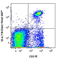
PE anti-mouse CD366 (Tim-3)

C57BL/6 mouse splenocytes were stained with anti-mouse CD3ε ... -
APC anti-mouse CD366 (Tim-3)

Mouse Tim-3 transfected cells were stained with anti-mouse C... -
PE/Cyanine7 anti-mouse CD366 (Tim-3)

Mouse Tim-3 transfected cells were stained with anti-mouse C... -
Biotin anti-mouse CD366 (Tim-3)

Mouse Tim-3 transfected cells were stained with biotinylated... -
PerCP/Cyanine5.5 anti-mouse CD366 (Tim-3)

Mouse Tim-3 transfected cells were stained with CD366 (Clone... -
GoInVivo™ Purified anti-mouse CD366 (Tim-3)

Mouse Tim-3 binds human Galectin-9 in a dose dependent manne... -
Brilliant Violet 605™ anti-mouse CD366 (Tim-3)

C57BL/6 mouse splenocytes were stained with anti-mouse CD3ε ... -
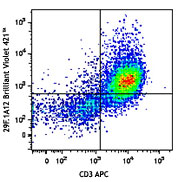
Brilliant Violet 421™ anti-mouse CD366 (Tim-3)

Mouse Tim-3 transfected cells were stained with anti-mouse C... -
Brilliant Violet 711™ anti-mouse CD366 (Tim-3)

C57BL/6 mouse splenocytes were stained with anti-mouse CD3ε ... -
Brilliant Violet 785™ anti-mouse CD366 (Tim-3)

Mouse Tim-3 transfected cells were stained with anti-mouse ... -
TotalSeq™-A0003 anti-mouse CD366 (Tim-3)
-
Ultra-LEAF™ Purified anti-mouse CD366 (Tim-3)

Mouse Tim-3 transfected cells were stained with LEAF™ purifi... -
APC/Fire™ 750 anti-mouse CD366 (Tim-3)

Mouse Tim-3 transfected cells were stained with anti-mouse C... -
TotalSeq™-C0003 anti-mouse CD366 (Tim-3)
-
TotalSeq™-B0003 anti-mouse CD366 (Tim-3)
-
Alexa Fluor® 647 anti-mouse CD366 (Tim-3)

Mouse Tim-3 transfected cells were stained with anti-mouse C... -
PE/Fire™ 810 anti-mouse CD366 (Tim-3)

Mouse Tim-3 transfected cells were stained with anti-mouse C... -
PE/Dazzle™ 594 anti-mouse CD366 (Tim-3)

Mouse Tim-3 transfected cells stained with anti-mouse CD366 ... -
PE/Fire™ 640 anti-mouse CD366 (Tim-3)

Mouse Tim-3 transfected cells were stained with anti-mouse C... -
KIRAVIA Blue 520™ anti-mouse CD366 (Tim-3)

Mouse Tim-3 transfected cells stained with anti-mouse CD366 ...


Follow Us