 Login/Register


- Clone
- 581 (See other available formats)
- Regulatory Status
- RUO
- Workshop
- V MA27
- Other Names
- Gp105-120, My10
- Isotype
- Mouse IgG1, κ
- Ave. Rating
- Submit a Review
- Product Citations
- publications

-

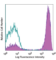
Human peripheral blood mononuclear cells were stained with CD45 FITC and CD34 Brilliant Violet 510™ (clone 581) or mouse IgG1, κ Brilliant Violet 510™ isotype control. Data was generated by gating on live CD14 negative cell population. -

| Cat # | Size | Price | Quantity Check Availability | Save | ||
|---|---|---|---|---|---|---|
| 343527 | 25 tests | $193 | ||||
| 343528 | 100 tests | $424 | ||||
CD34, also known as gp105-120, is a type I monomeric sialomucin-like glycophosphoprotein with an approximate molecular weight of 105-120 kD. Selectively expressed on the majority of hematopoietic stem/progenitor cells, bone marrow stromal cells, capillary endothelial cells, embryonic fibroblasts, and some nervous tissue, CD34 is a commonly used marker to identify human hematopoietic stem/progenitor cells. According to the differential sensitivity to enzymatic cleavage, four groups of epitopes of CD34 have been described. CD34 mediates cell adhesion and lymphocytes homing through binding to L-selectin and E-selectin ligands.
Product DetailsProduct Details
- Verified Reactivity
- Human
- Reported Reactivity
- Cynomolgus
- Antibody Type
- Monoclonal
- Host Species
- Mouse
- Formulation
- Phosphate-buffered solution, pH 7.2, containing 0.09% sodium azide and BSA (origin USA).
- Preparation
- The antibody was purified by affinity chromatography and conjugated with Brilliant Violet 510™ under optimal conditions.
- Concentration
- Lot-specific (to obtain lot-specific concentration and expiration, please enter the lot number in our Certificate of Analysis online tool.)
- Storage & Handling
- The antibody solution should be stored undiluted between 2°C and 8°C, and protected from prolonged exposure to light. Do not freeze.
- Application
-
FC - Quality tested
- Recommended Usage
-
Each lot of this antibody is quality control tested by immunofluorescent staining with flow cytometric analysis. For flow cytometric staining, the suggested use of this reagent is 5 µl per million cells in 100 µl staining volume or 5 µl per 100 µl of whole blood.
Brilliant Violet 510™ excites at 405 nm and emits at 510 nm. The bandpass filter 510/50 nm is recommended for detection, although filter optimization may be required depending on other fluorophores used. Be sure to verify that your cytometer configuration and software setup are appropriate for detecting this channel. Refer to your instrument manual or manufacturer for support. Brilliant Violet 510™ is a trademark of Sirigen Group Ltd.
Learn more about Brilliant Violet™.
This product is subject to proprietary rights of Sirigen Inc. and is made and sold under license from Sirigen Inc. The purchase of this product conveys to the buyer a non-transferable right to use the purchased product for research purposes only. This product may not be resold or incorporated in any manner into another product for resale. Any use for therapeutics or diagnostics is strictly prohibited. This product is covered by U.S. Patent(s), pending patent applications and foreign equivalents. - Excitation Laser
-
Violet Laser (405 nm)
- Application Notes
-
The 581 antibody recognizes the class III group epitope which is resistant to sialidase/glycolyprotease and chymopapain treatment. Additional reported applications (for the relevant formats) include: immunohistochemical staining of paraffin-embedded tissue sections5 and immunofluorescence6.
-
Application References
(PubMed link indicates BioLegend citation) -
- Schlossman SF, et al. 1995. Leukocyte Typing V:White Cell Differentiation Antigen. New York:Oxford University Press.
- Felschow DM, et al. 2001. Blood 97:3768.
- Rudin CE, et al. 1997. Br. J. Haematol. 97:488.
- Yoshino N, et al. 2000. Exp. Anim. (Tokyo) 49:97. (FC)
- Skowasch D, et al. 2003. Cardiovasc Res. 60:684. (IHC)
- Umland O, et al. 2003. J. Histochem. Cytochem. 51:977. (IF)
- Product Citations
-
- RRID
-
AB_2561985 (BioLegend Cat. No. 343527)
AB_2563856 (BioLegend Cat. No. 343528)
Antigen Details
- Structure
- 105-120 kD single chain mucin-like glycoprotein
- Distribution
-
Hematopoietic stem/progenitor cells, bone marrow stromal cells, endothelial cells, embryonic fibroblasts
- Function
- Cell adhesion
- Ligand/Receptor
- L-selectin, E-selectin
- Cell Type
- Endothelial cells, Fibroblasts, Hematopoietic stem and progenitors
- Biology Area
- Cell Biology, Immunology, Neuroinflammation, Neuroscience, Stem Cells
- Molecular Family
- CD Molecules
- Antigen References
-
1. Krause DS, et al. 1996. Blood 87:1.
2. Puri KD, et al. 1995. J. Cell Biol. 131:261.
3. Zola H, et al. 2007. Leukocyte and Stromal Cell Molecules:The CD Markers. John Wiley & Sons Inc, Hoboken New Jersey. - Gene ID
- 947 View all products for this Gene ID
- UniProt
- View information about CD34 on UniProt.org
Related Pages & Pathways
Pathways


Other Formats
View All CD34 Reagents Request Custom Conjugation| Description | Clone | Applications |
|---|---|---|
| Purified anti-human CD34 | 581 | FC,CyTOF®,ICC,IHC-P |
| FITC anti-human CD34 | 581 | FC |
| PE anti-human CD34 | 581 | FC |
| Alexa Fluor® 647 anti-human CD34 | 581 | FC |
| APC anti-human CD34 | 581 | FC |
| Pacific Blue™ anti-human CD34 | 581 | FC |
| APC/Cyanine7 anti-human CD34 | 581 | FC |
| PE/Cyanine7 anti-human CD34 | 581 | FC |
| Alexa Fluor® 488 anti-human CD34 | 581 | FC |
| PerCP anti-human CD34 | 581 | FC |
| PerCP/Cyanine5.5 anti-human CD34 | 581 | FC |
| Biotin anti-human CD34 | 581 | FC |
| Alexa Fluor® 700 anti-human CD34 | 581 | FC |
| Brilliant Violet 510™ anti-human CD34 | 581 | FC |
| Purified anti-human CD34 (Maxpar® Ready) | 581 | FC,CyTOF® |
| PE/Dazzle™ 594 anti-human CD34 | 581 | FC |
| APC/Fire™ 750 anti-human CD34 | 581 | FC |
| TotalSeq™-A0054 anti-human CD34 | 581 | PG |
| TotalSeq™-B0054 anti-human CD34 | 581 | PG |
| TotalSeq™-C0054 anti-human CD34 | 581 | PG |
| TotalSeq™-D0054 anti-human CD34 | 581 | PG |
| Spark Red™ 718 anti-human CD34 | 581 | FC |
| Cell-Vive™ GMP Ultra-LEAF™ Purified anti-human CD34 SF | 581 | FC,ICC,IHC-P,CyTOF® |
| Spark PLUS UV™ 395 anti-human CD34 | 581 | FC |
Customers Also Purchased
Compare Data Across All Formats
This data display is provided for general comparisons between formats.
Your actual data may vary due to variations in samples, target cells, instruments and their settings, staining conditions, and other factors.
If you need assistance with selecting the best format contact our expert technical support team.
-
Purified anti-human CD34

Human myeloid cell line KG1a stained with purified 581, foll... -
FITC anti-human CD34

Human peripheral blood mononuclear cells stained with 581 FI... 
-
PE anti-human CD34

Human peripheral blood mononuclear cells stained with 581 PE... 
-
Alexa Fluor® 647 anti-human CD34

Human peripheral blood leukocytes stained with 581 Alexa Flu... 
-
APC anti-human CD34

Human peripheral blood leukocytes stained with 581 APC/CD45 ... 
-
Pacific Blue™ anti-human CD34

Human peripheral blood leukocytes stained with CD34 Pacific... 
-
APC/Cyanine7 anti-human CD34

Human peripheral blood mononuclear cells were stained with C... -
PE/Cyanine7 anti-human CD34

Human peripheral blood stained with 581 PE/Cyanine7 or mouse... 
-
Alexa Fluor® 488 anti-human CD34

Human peripheral blood leukocytes stained with CD45 (HI30) A... 
-
PerCP anti-human CD34

Human peripheral blood leukocytes stained with CD45 FITC and... 
-
PerCP/Cyanine5.5 anti-human CD34

Human peripheral blood was stained with CD45 APC and CD34 (C... -
Biotin anti-human CD34

Human peripheral blood lymphocytes stained with biotinylated... 
-
Alexa Fluor® 700 anti-human CD34

Human peripheral blood mononuclear cells were stained with C... 
-
Brilliant Violet 510™ anti-human CD34

Human peripheral blood mononuclear cells were stained with C... 
-
Purified anti-human CD34 (Maxpar® Ready)

Human KG-1a cells (blue) and mouse EL4 T cells (red) stained... -
PE/Dazzle™ 594 anti-human CD34

Human peripheral blood leukocytes were stained with CD14 Bri... 
-
APC/Fire™ 750 anti-human CD34

Human peripheral blood mononuclear cells were stained with C... 
-
TotalSeq™-A0054 anti-human CD34
-
TotalSeq™-B0054 anti-human CD34
-
TotalSeq™-C0054 anti-human CD34
-
TotalSeq™-D0054 anti-human CD34
-
Spark Red™ 718 anti-human CD34

Human peripheral blood leukocytes were stained with anti-hum... -
Cell-Vive™ GMP Ultra-LEAF™ Purified anti-human CD34 SF

Human myeloid cell line KG1a stained with Cell-Vive™ GMP Ult... -
Spark PLUS UV™ 395 anti-human CD34

Human peripheral blood leukocytes were stained with anti-hum...


Follow Us