 Login/Register


- Clone
- DIH9 (See other available formats)
- Regulatory Status
- RUO
- Other Names
- T1/ST2, ST2L, IL-1R1, T1, DER4, IL1R1
- Isotype
- Rat IgG2a, κ
- Ave. Rating
- Submit a Review
- Product Citations
- publications

-

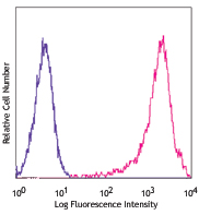
Mouse Th2 clone D10.G4.1 was stained with anti-mouse IL-33Ra/ST2 (clone DIH9) APC (filled histogram) or rat IgG1, κ APC isotype control (open histogram).
| Cat # | Size | Price | Quantity Check Availability | Save | ||
|---|---|---|---|---|---|---|
| 145305 | 25 µg | $161 | ||||
| 145306 | 100 µg | $362 | ||||
IL-33Rα, also known as ST2 or IL-1RL1, is a member of the Toll/IL-1 receptor family. It binds IL-33 and is structurally similar to IL-1R1. Two forms of the protein exist - a soluble form known as ST2 and a membrane anchored form known as ST2L. The membrane form is expressed by Th2 cells and bone marrow derived mast cells, whereas the soluble form is expressed by serum-stimulated fibroblasts.
Blocking with anti-ST2 antibodies has been shown to alleviate experimental arthritis and airway inflammation. The IL-33-ST2 axis has been reported to be important across a range of diseases including asthma, allergies, and cardiac disease.
Product Details
- Verified Reactivity
- Mouse
- Antibody Type
- Monoclonal
- Host Species
- Rat
- Immunogen
- IL-33Rα-hFcγ1 fusion protein.
- Formulation
- Phosphate-buffered solution, pH 7.2, containing 0.09% sodium azide.
- Preparation
- The antibody was purified by affinity chromatography and conjugated with APC under optimal conditions.
- Concentration
- 0.2 mg/ml
- Storage & Handling
- The antibody solution should be stored undiluted between 2°C and 8°C, and protected from prolonged exposure to light. Do not freeze.
- Application
-
FC - Quality tested
- Recommended Usage
-
Each lot of this antibody is quality control tested by immunofluorescent staining with flow cytometric analysis. For flow cytometric staining, the suggested use of this reagent is ≤1.0 µg per million cells in 100 µl volume. It is recommended that the reagent be titrated for optimal performance for each application.
- Excitation Laser
-
Red Laser (633 nm)
-
Application References
(PubMed link indicates BioLegend citation) -
- Hashiguchi M, et al. 2014. Eur. J. Immunology. (FC) PubMed
- Product Citations
-
- RRID
-
AB_2561916 (BioLegend Cat. No. 145305)
AB_2561917 (BioLegend Cat. No. 145306)
Antigen Details
- Structure
- 63 kD, IL-1 receptor family member, single pass type I membrane protein
- Distribution
-
Mast cells, Th2 cells, and fibroblasts
- Function
- Following interaction with IL-33, it recruits MyD88, Irak1, Irak4 and Traf6 followed by phosphorylation of the MAP/ERK pathway.
- Interaction
- Interacts with MyD88, Irak1, Irak4 and Traf6
- Ligand/Receptor
- IL-33
- Cell Type
- Fibroblasts, Mast cells, Th2
- Biology Area
- Immunology, Innate Immunity
- Molecular Family
- Cytokine/Chemokine Receptors
- Antigen References
-
1. Yanagisawa K, et al. 1993. FEBS Lett. 318:83.
2. Schmitt E, et al. 1990. Cytokine 6:407.
3. Yanagisawa K, et al. 1992. FEBS Lett. 302:51.
4. Takagi T, et al. 1993. Biochim Biophys Acta. 1178:194. - Gene ID
- 17082 View all products for this Gene ID
- UniProt
- View information about IL-33Ralpha on UniProt.org
Related Pages & Pathways
Pathways


Related FAQs
Other Formats
View All IL-33Rα Reagents Request Custom ConjugationCustomers Also Purchased
Compare Data Across All Formats
This data display is provided for general comparisons between formats.
Your actual data may vary due to variations in samples, target cells, instruments and their settings, staining conditions, and other factors.
If you need assistance with selecting the best format contact our expert technical support team.
-
Purified anti-mouse IL-33Rα (IL1RL1, ST2)

Mouse Th2 clone D10.G4.1 cell line was stained with purified... -
PE anti-mouse IL-33Rα (IL1RL1, ST2)

Mouse Th2 clone D10.G4.1 was stained with anti-mouse IL-33Ra... -
APC anti-mouse IL-33Rα (IL1RL1, ST2)

Mouse Th2 clone D10.G4.1 was stained with anti-mouse IL-33Ra... -
PerCP/Cyanine5.5 anti-mouse IL-33Rα (IL1RL1, ST2)

ST2-FLAG transfected DO-11.10 mouse cells were stained with ... -
Brilliant Violet 421™ anti-mouse IL-33Rα (IL1RL1, ST2)

Mouse Th2 clone D10.G4.1 was stained with anti-mouse IL-33Ra... -
Biotin anti-mouse IL-33Rα (IL1RL1, ST2)

Mouse Th2 clone D10.G4.1 was stained with biotinylated anti-... -
PE/Dazzle™ 594 anti-mouse IL-33Rα (IL1RL1, ST2)

Mouse Th2 clone D10.G4.1 was stained with anti-mouse IL-33Ra... -
PE/Cyanine7 anti-mouse IL-33Rα (IL1RL1, ST2)

Mouse Th2 clone D10.G4.1 was stained with anti-mouse IL-33Ra... -
TotalSeq™-A0837 anti-mouse IL-33Rα (IL1RL1, ST2)
-
TotalSeq™-C0837 anti-mouse IL-33Rα (IL1RL1, ST2)
-
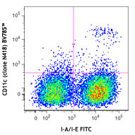
Brilliant Violet 785™ anti-mouse IL-33Rα (IL1RL1, ST2)

Mouse Th2 clone D10.G4.1 was stained with anti-mouse IL-33Rα... -
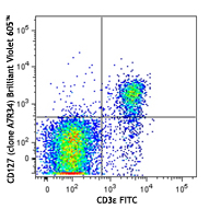
Brilliant Violet 605™ anti-mouse IL-33Rα (IL1RL1, ST2)

Mouse Th2 clone D10.G4.1 was stained with anti-mouse IL-33Rα... -
APC/Fire™ 750 anti-mouse IL-33Rα (IL1RL1, ST2)

Mouse Th2 clone D10.G4.1 was stained with anti-mouse IL-33Rα... -
Brilliant Violet 711™ anti-mouse IL-33Rα (IL1RL1, ST2)

Mouse ST2-Flag transfected D011.10 cells were stained with a... -
TotalSeq™-B0837 anti-mouse IL-33Rα (IL1RL1, ST2)


Follow Us