 Login/Register


- Clone
- 11-26c.2a (See other available formats)
- Regulatory Status
- RUO
- Other Names
- Immunoglobulin D
- Isotype
- Rat IgG2a, κ
- Ave. Rating
- Submit a Review
- Product Citations
- publications

-

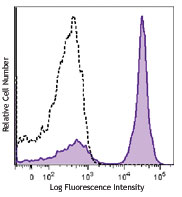
C57BL/6 mouse splenocytes were stained with B220 FITC and IgD (clone 11-26c.2a) Alexa Fluor® 700 (top) or rat IgG2a, κ Alexa Fluor® 700 isotype control (bottom). -

-

Confocal image of C57BL/6 mouse spleen sample acquired using the IBEX method of highly multiplexed antibody-based imaging: CD4 (cyan), CD8 (magenta), and IgD (blue) in Cycle 1. Tissues were prepared using ~1% (vol/vol) formaldehyde and a detergent. Following fixation, samples are immersed in 30% (wt/vol) sucrose for cryoprotection. Images are courtesy of Drs. Andrea J. Radtke and Ronald N. Germain of the Center for Advanced Tissue Imaging (CAT-I) in the National Institute of Allergy and Infectious Diseases (NIAID, NIH). -

Confocal image of C57BL/6 mouse small intestine sample acquired using the IBEX method of highly multiplexed antibody-based imaging: EpCAM (blue) in Cycle 1, IgD (red) in Cycle 1, and CD11c (cyan) in Cycle 3. Tissues were prepared using ~1% (vol/vol) formaldehyde and a detergent. Following fixation, samples are immersed in 30% (wt/vol) sucrose for cryoprotection. Images are courtesy of Drs. Andrea J. Radtke and Ronald N. Germain of the Center for Advanced Tissue Imaging (CAT-I) in the National Institute of Allergy and Infectious Diseases (NIAID, NIH).
| Cat # | Size | Price | Quantity Check Availability | Save | ||
|---|---|---|---|---|---|---|
| 405729 | 25 µg | $124 | ||||
| 405730 | 100 µg | $293 | ||||
Product Details
- Verified Reactivity
- Mouse
- Antibody Type
- Monoclonal
- Host Species
- Rat
- Formulation
- Phosphate-buffered solution, pH 7.2, containing 0.09% sodium azide.
- Preparation
- The antibody was purified by affinity chromatography and conjugated with Alexa Fluor® 700 under optimal conditions.
- Concentration
- 0.5 mg/ml
- Storage & Handling
- The antibody solution should be stored undiluted between 2°C and 8°C, and protected from prolonged exposure to light. Do not freeze.
- Application
-
FC - Quality tested
SB - Reported in the literature, not verified in house
- Recommended Usage
-
Each lot of this antibody is quality control tested by immunofluorescent staining with flow cytometric analysis. For flow cytometric staining, the suggested use of this reagent is ≤0.25 µg per million cells in 100 µl volume. It is recommended that the reagent be titrated for optimal performance for each application.
* Alexa Fluor® 700 has a maximum emission of 719 nm when it is excited at 633 nm / 635 nm. Prior to using Alexa Fluor® 700 conjugate for flow cytometric analysis, please verify your flow cytometer's capability of exciting and detecting the fluorochrome.
Alexa Fluor® and Pacific Blue™ are trademarks of Life Technologies Corporation.
View full statement regarding label licenses - Excitation Laser
-
Red Laser (633 nm)
- Application Notes
-
The 11-26c.2a antibody reacts with immunoglobulin D in all tested mouse haplotypes. The antibody binds membrane IgD expressed on most B cells. The 11-26c.2a antibody neither induces proliferation of splenic B cells nor induces B cell activation. Additional reported applications (for the relevant formats) include: immunohistochemical staining of acetone-fixed frozen sections2,3, and spatial biology (IBEX)10,11.
- Additional Product Notes
-
Iterative Bleaching Extended multi-pleXity (IBEX) is a fluorescent imaging technique capable of highly-multiplexed spatial analysis. The method relies on cyclical bleaching of panels of fluorescent antibodies in order to image and analyze many markers over multiple cycles of staining, imaging, and, bleaching. It is a community-developed open-access method developed by the Center for Advanced Tissue Imaging (CAT-I) in the National Institute of Allergy and Infectious Diseases (NIAID, NIH).
-
Application References
(PubMed link indicates BioLegend citation) -
- Nitschke L, et al. 1993. P. Natl. Acad. Sci. USA 90:1887. (FC)
- Weih D, et al. 2001. J. Immunol. 167:1909. (IHC)
- Koni PA, et al. 2001. J. Exp. Med. 193:741. (IHC)
- Ahuja A, et al. 2007. J. Immunol. 179:3351. (FC) PubMed
- Haynes NM, et al. 2007. J. Immunol. 179:5099. (FC)
- Good-Jacobson KL, et al. 2010. Nat. Immunol. 11:535. (FC) PubMed
- Tomayko MM, et al. 2010. J. Immunol. 185:7146. PubMed
- Park SY, et al. 2013. J. Immunol. 190:1094. PubMed
- Rouaud P, et al. 2014. J Exp Med. 211:975. PubMed
- Radtke AJ, et al. 2020. Proc Natl Acad Sci U S A. 117:33455-65. (SB) PubMed
- Radtke AJ, et al. 2022. Nat Protoc. 17:378-401. (SB) PubMed
- Product Citations
-
- RRID
-
AB_2563340 (BioLegend Cat. No. 405729)
AB_2563341 (BioLegend Cat. No. 405730)
Antigen Details
- Structure
- Ig family
- Distribution
-
B cells
- Function
- B cell differentiation
- Cell Type
- B cells
- Biology Area
- Immunology
- Gene ID
- 380797 View all products for this Gene ID
- UniProt
- View information about IgD on UniProt.org
Related Pages & Pathways
Pathways


Related FAQs
- If an antibody clone has been previously successfully used in IBEX in one fluorescent format, will other antibody formats work as well?
-
It’s likely that other fluorophore conjugates to the same antibody clone will also be compatible with IBEX using the same sample fixation procedure. Ultimately a directly conjugated antibody’s utility in fluorescent imaging and IBEX may be specific to the sample and microscope being used in the experiment. Some antibody clone conjugates may perform better than others due to performance differences in non-specific binding, fluorophore brightness, and other biochemical properties unique to that conjugate.
- Will antibodies my lab is already using for fluorescent or chromogenic IHC work in IBEX?
-
Fundamentally, IBEX as a technique that works much in the same way as single antibody panels or single marker IF/IHC. If you’re already successfully using an antibody clone on a sample of interest, it is likely that clone will have utility in IBEX. It is expected some optimization and testing of different antibody fluorophore conjugates will be required to find a suitable format; however, legacy microscopy techniques like chromogenic IHC on fixed or frozen tissue is an excellent place to start looking for useful antibodies.
- Are other fluorophores compatible with IBEX?
-
Over 18 fluorescent formats have been screened for use in IBEX, however, it is likely that other fluorophores are able to be rapidly bleached in IBEX. If a fluorophore format is already suitable for your imaging platform it can be tested for compatibility in IBEX.
- The same antibody works in one tissue type but not another. What is happening?
-
Differences in tissue properties may impact both the ability of an antibody to bind its target specifically and impact the ability of a specific fluorophore conjugate to overcome the background fluorescent signal in a given tissue. Secondary stains, as well as testing multiple fluorescent conjugates of the same clone, may help to troubleshoot challenging targets or tissues. Using a reference control tissue may also give confidence in the specificity of your staining.
- How can I be sure the staining I’m seeing in my tissue is real?
-
In general, best practices for validating an antibody in traditional chromogenic or fluorescent IHC are applicable to IBEX. Please reference the Nature Methods review on antibody based multiplexed imaging for resources on validating antibodies for IBEX.
Other Formats
View All IgD Reagents Request Custom ConjugationCustomers Also Purchased
Compare Data Across All Formats
This data display is provided for general comparisons between formats.
Your actual data may vary due to variations in samples, target cells, instruments and their settings, staining conditions, and other factors.
If you need assistance with selecting the best format contact our expert technical support team.
-
FITC anti-mouse IgD

C57BL/6 mouse splenocytes stained with 11-26c.2a FITC -
PE anti-mouse IgD

C57BL/6 splenocytes stained with 11-26c.2a PE -
Purified anti-mouse IgD

C57BL/6 mouse splenocytes stained with purified 11-26c.2a, f... -
PerCP anti-mouse IgD

C57BL/6 mouse splenocytes were stained with CD45R/B220 APC a... 
-
Biotin anti-mouse IgD

C57BL/6 splenocytes were stained with CD45R/B220 APC and bio... 
-
Brilliant Violet 711™ anti-mouse IgD

C57BL/6 mouse splenocytes were stained with B220 APC and IgD... 
-
Alexa Fluor® 700 anti-mouse IgD

C57BL/6 mouse splenocytes were stained with B220 FITC and Ig... 

Confocal image of C57BL/6 mouse spleen sample acquired using... 
Confocal image of C57BL/6 mouse small intestine sample acqui... -
Alexa Fluor® 647 anti-mouse IgD

C57BL/6 splenocytes stained with 11-26c.2a Alexa Fluor® 647 
Paraformaldehyde-fixed (4%), 500 μm-thick mouse spleen secti... -
PerCP/Cyanine5.5 anti-mouse IgD

C57BL/6 splenocytes stained with 11-26c.2a PerCP/Cyanine5.5 -
Pacific Blue™ anti-mouse IgD

C57BL/6 splenocytes stained with 11-26c.2a Pacific Blue&trad... 
C57BL/6 splenocytes stained with rat IgG2a Pacific Blue&trad... -
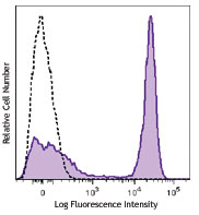
APC anti-mouse IgD

C57BL/6 splenocytes stained with 11-26c.2a APC -
APC/Cyanine7 anti-mouse IgD

C57BL/6 splenocytes stained with 11-26c.2a APC/Cyanine7 -
Alexa Fluor® 488 anti-mouse IgD

C57BL/6 splenocytes were stained with B220 APC and IgD (11-2... 

Mice were injected subcutaneously with sheep red blood cells... -
PE/Cyanine7 anti-mouse IgD

C57BL/6 splenocytes were stained with CD45R/B220 FITC and Ig... -
Brilliant Violet 650™ anti-mouse IgD

C57BL/6 splenocytes were stained with CD45R/B220 APC and IgD... 
-
Brilliant Violet 510™ anti-mouse IgD

C57BL/6 splenocytes were stained with CD45R/B220 APC and IgD... 
-
Brilliant Violet 421™ anti-mouse IgD

C57BL/6 splenocytes were stained with CD45R/B220 APC and IgD... 
-
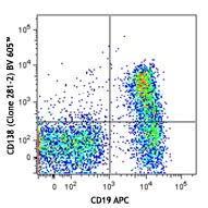
Brilliant Violet 605™ anti-mouse IgD

C57BL/6 splenocytes were stained with CD45R/B220 APC and IgD... 
-
Purified anti-mouse IgD (Maxpar® Ready)

C57BL/6 mouse splenocytes stained with 176Yb-anti-CD45R/B220... -
Alexa Fluor® 594 anti-mouse IgD

C57BL/6 mouse frozen spleen section was fixed with 4% parafo... 
Paraformaldehyde-fixed (4%), 500 μm-thick mouse spleen secti... -
PE/Dazzle™ 594 anti-mouse IgD

C57BL/6 mouse splenocytes were stained with B220 APC and IgD... 
-
APC/Fire™ 750 anti-mouse IgD

C57BL/6 mouse splenocytes were stained with CD45R/B220 FITC ... 
-
TotalSeq™-A0571 anti-mouse IgD
-
TotalSeq™-C0571 anti-mouse IgD
-
Spark NIR™ 685 anti-mouse IgD

C57BL/6 splenocytes were stained with anti-mouse CD45R/B220 ... -
TotalSeq™-B0571 anti-mouse IgD Antibody
-
Spark Violet™ 423 anti-mouse IgD

C57BL/6 splenocytes were stained with anti-mouse/human CD45R... -
PE/Cyanine5 anti-mouse IgD

C57BL/6 mouse splenocytes were stained with anti-mouse B220 ... -
Brilliant Violet 785™ anti-mouse IgD

C57BL/6 mouse splenocytes were stained with anti-mouse B220 ...


Follow Us