 Login/Register


- Clone
- 3D6.112 (See other available formats)
- Regulatory Status
- RUO
- Other Names
- Sialic acid binding Ig-like lectin 1 (Siglec-1), Sialoadhesin (Sn)
- Isotype
- Rat IgG2a, κ
- Ave. Rating
- Submit a Review
- Product Citations
- publications

-

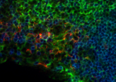
C57BL/6 mouse frozen spleen section was fixed with 4% paraformaldehyde (PFA) for 10 minutes at room temperature and blocked with 5% FBS plus 5% rat serum for 30 minutes at room temperature. Then the section was stained with 5 µg/ml of CD169 (clone 3D6.112) Alexa Fluor® 594 (red), B220 (blue), and CD3 (green) overnight at 4°C. The image was captured with a 10X objective. -

Paraformaldehyde-fixed (4%), 500 µm-thick mouse spleen section was processed according to the Ce3D™ Tissue Clearing Kit protocol (Cat. No. 427701). The section was costained with anti-mouse CD169 (Siglec-1) Antibody (clone 3D6.112) Alexa Fluor® 594 at 5 µg/mL (yellow), and anti-mouse/human CD11b Antibody (clone M1/70) Alexa Fluor® 647 at 5 µg/mL (magenta). The section was then optically cleared and mounted in a sample chamber. The image was captured with a 10X objective using Zeiss 780 confocal microscope and processed by Imaris image analysis software.
Watch the video.
| Cat # | Size | Price | Quantity Check Availability | Save | ||
|---|---|---|---|---|---|---|
| 142416 | 100 µg | $382 | ||||
CD169, also known as Siglec-1 and Sialoadhesin (Sn), is a type I lectin containing 17 immunoglobulin (Ig) domains (one variable domain and 16 constant domains). CD169 binds to sialic acids, which can be found on PSGL-1, CD43, CD206, and CD227. By its affinity to α2, 3-linked sialic acid, it is involved in macrophage binding to different cell types such as granulocytes, monocytes, NK, B, and T cells. CD169 was initially identified as a sialic acid-dependent sheep erythrocyte receptor (SER) on resident bone marrow cells of mice. It has been identified as highly expressed on resident bone marrow macrophages which plays an important role in retention of stem cells in mesenchymal stem cell niche. It is also found on some specific subsets of tissue macrophages in spleen, lymph nodes, bone marrow, liver, colon, lungs, and cancer cells. Evidence suggest that CD169-positive macrophages serve as lymph node-resident APCs to dominate early activation of tumor antigen-specific CD8+ T cells and invariant NK cell.
Product DetailsProduct Details
- Verified Reactivity
- Mouse
- Antibody Type
- Monoclonal
- Host Species
- Rat
- Immunogen
- Purified Native Sialoadhesin from spleen
- Formulation
- Phosphate-buffered solution, pH 7.2, containing 0.09% sodium azide.
- Preparation
- The antibody was purified by affinity chromatography and conjugated with Alexa Fluor® 594 under optimal conditions.
- Concentration
- 0.5 mg/ml
- Storage & Handling
- The antibody solution should be stored undiluted between 2°C and 8°C, and protected from prolonged exposure to light. Do not freeze.
- Application
-
IHC-F - Quality tested
3D IHC - Verified - Recommended Usage
-
Each lot of this antibody is quality control tested by immunohistochemical staining on frozen tissue sections. For immunohistochemistry, a concentration range of 2.5 - 10 µg/ml is suggested. For 3D immunohistochemistry on formalin-fixed tissues, a concentration of 5.0 µg/mL is suggested. It is recommended that the reagent be titrated for optimal performance for each application.
* Alexa Fluor® 594 has an excitation maximum of 590 nm, and a maximum emission of 617 nm.
Alexa Fluor® and Pacific Blue™ are trademarks of Life Technologies Corporation.
View full statement regarding label licenses - Application Notes
-
Additional reported applications (for the relevant formats) include: immunohistochemical staining in frozen tissue sections1, and spatial biology (IBEX)4,5.
-
Application References
(PubMed link indicates BioLegend citation) - Product Citations
-
- RRID
-
AB_2565620 (BioLegend Cat. No. 142416)
Antigen Details
- Structure
- Type I single membrane-spanning lectin containing 17 immunoglobulin (Ig) domains, belongs to the immunoglobulin superfamily.
- Distribution
-
Macrophages in spleen, lymph nodes, bone marrow, liver, colon and lungs.
- Function
- Adhesion.
- Ligand/Receptor
- PSGL-1, CD43, CD206 and CD227.
- Cell Type
- Macrophages
- Biology Area
- Cell Biology, Immunology
- Molecular Family
- Adhesion Molecules, CD Molecules, Protein Kinases/Phosphatase, Siglec Molecules
- Antigen References
-
1. Chow A, et al. 2011. J. Exp. Med. 208:261.
2. Asano K, et al. 2011. Immunity 34:85.
3. Xiong YS, et al. 2009. Clin. Biochem. 42:1057.
4. Varki A, et al. 2009. Glycoconj. J. 26:231.
5. Rempel H, et al. 2008. PLoS One 3:e1967.
6. Crocker PR, et al. 2001. Trends Immunol. 22:337.
7. Hartnell A, et al. 2001. Blood 97:288.
8. Crocker PR, et al. 1985. J. Exp. Med. 162:993. - Gene ID
- 20612 View all products for this Gene ID
- UniProt
- View information about CD169 on UniProt.org


Related FAQs
Other Formats
View All CD169 Reagents Request Custom Conjugation| Description | Clone | Applications |
|---|---|---|
| Purified anti-mouse CD169 (Siglec-1) | 3D6.112 | FC,IHC-F,SB |
| PE anti-mouse CD169 (Siglec-1) | 3D6.112 | FC,SB |
| FITC anti-mouse CD169 (Siglec-1) | 3D6.112 | FC |
| Alexa Fluor® 647 anti-mouse CD169 (Siglec-1) | 3D6.112 | FC,IHC-F,3D IHC |
| PerCP/Cyanine5.5 anti-mouse CD169 (Siglec-1) | 3D6.112 | FC |
| PE/Cyanine7 anti-mouse CD169 (Siglec-1) | 3D6.112 | FC |
| Brilliant Violet 605™ anti-mouse CD169 (Siglec-1) | 3D6.112 | FC |
| APC anti-mouse CD169 (Siglec-1) | 3D6.112 | FC |
| Alexa Fluor® 594 anti-mouse CD169 (Siglec-1) | 3D6.112 | IHC-F,3D IHC |
| Alexa Fluor® 488 anti-mouse CD169 (Siglec-1) | 3D6.112 | IHC-F,3D IHC |
| Brilliant Violet 421™ anti-mouse CD169 (Siglec-1) | 3D6.112 | IHC-F |
| PE/Dazzle™ 594 anti-mouse CD169 (Siglec-1) | 3D6.112 | FC |
| TotalSeq™-A0440 anti-mouse CD169 (Siglec-1) | 3D6.112 | PG |
| TotalSeq™-C0440 anti-mouse CD169 (Siglec-1) | 3D6.112 | PG |
| TotalSeq™-B0440 anti-mouse CD169 (Siglec-1) | 3D6.112 | PG |
| Biotin anti-mouse CD169 (Siglec-1) | 3D6.112 | FC |
Customers Also Purchased
Compare Data Across All Formats
This data display is provided for general comparisons between formats.
Your actual data may vary due to variations in samples, target cells, instruments and their settings, staining conditions, and other factors.
If you need assistance with selecting the best format contact our expert technical support team.
-
Purified anti-mouse CD169 (Siglec-1)

C57BL/6 splenocytes were stained with F4/80 APC, Ly-6G PerCP... 

Fresh, frozen mouse spleen was stained with purified CD169 c... -
PE anti-mouse CD169 (Siglec-1)
C57BL/6 splenocytes were stained with F4/80 APC, Ly-6G PerCP...

C57BL/6 inguinal lymph node labeled in-vivo with PE anti-mou... 
Live intravital mouse spleen imaging. PE CD169 (red) (clone ... 
Fixed whole mount mouse spleen imaging sectioned after intra... 
Confocal image of C57BL/6 mouse spleen sample acquired using... -
FITC anti-mouse CD169 (Siglec-1)

C57BL/6 mouse bone marrow cells were stained with F4/80 PE, ... 
-
Alexa Fluor® 647 anti-mouse CD169 (Siglec-1)

C57BL/6 mouse bone marrow cells were stained with F4/80 PE, ... 

C57BL/6 mouse frozen lymph node section was fixed with 4% pa... Formalin-fixed, 300 micron-thick mouse spleen section was bl... 
Paraformaldehyde-fixed (1%), 500 μm-thick mouse spleen secti... -
PerCP/Cyanine5.5 anti-mouse CD169 (Siglec-1)

C57BL/6 mouse bone marrow cells were stained with Ly-6G APC ... -
PE/Cyanine7 anti-mouse CD169 (Siglec-1)

C57BL/6 mouse bone marrow cells were stained with Ly-6G FITC... -
Brilliant Violet 605™ anti-mouse CD169 (Siglec-1)

C57BL/6 mouse bone marrow cells were stained with Ly-6G FITC... 
-
APC anti-mouse CD169 (Siglec-1)

C57BL/6 mouse bone marrow cells were stained with Ly-6G PE a... 
-
Alexa Fluor® 594 anti-mouse CD169 (Siglec-1)

C57BL/6 mouse frozen spleen section was fixed with 4% parafo... 
Paraformaldehyde-fixed (4%), 500 µm-thick mouse spleen secti... -
Alexa Fluor® 488 anti-mouse CD169 (Siglec-1)

C57BL/6 mouse frozen spleen section was fixed with 4% parafo... 
Dissected C57/B6 mouse spleen was immersed in 4% paraformal... 
Formalin-fixed, 400 micron-thick mouse spleen section was bl... -
Brilliant Violet 421™ anti-mouse CD169 (Siglec-1)

C57BL/6 mouse frozen spleen section was fixed with 4% parafo... -
PE/Dazzle™ 594 anti-mouse CD169 (Siglec-1)

C57BL/6 mouse bone marrow cells were stained with Ly-6G FITC... -
TotalSeq™-A0440 anti-mouse CD169 (Siglec-1)
-
TotalSeq™-C0440 anti-mouse CD169 (Siglec-1)
-
TotalSeq™-B0440 anti-mouse CD169 (Siglec-1)
-
Biotin anti-mouse CD169 (Siglec-1)

C57BL/6 mouse bone marrow cells were stained with anti-mouse...


Follow Us