 Login/Register


- Clone
- W19260A (See other available formats)
- Regulatory Status
- RUO
- Other Names
- Yes1 Associated Transcriptional Regulator, YAP65, Yes-Associated Protein YAP65 Homolog, Transcriptional Coactivator YAP1, Yes Associated Protein 1, COB1, YAP2, YAP, YKI
- Isotype
- Rat IgG2b, κ
- Ave. Rating
- Submit a Review
- Product Citations
- publications

-

Whole cell extracts (15 µg total protein) from HeLa, MCF7 (positive controls), Raji (negative control) and NIH/3T3 cells were resolved by 4-12% Bis-Tris gel electrophoresis, transferred to a PVDF membrane, and probed with 1.0 μg/mL (1:500 dilution) purified anti-YAP1 (clone W19260A) overnight at 4°C. Proteins were visualized by chemiluminescence detection using HRP goat anti-rat IgG (Cat. No. 405405) at a 1:3000 dilution. Western-Ready™ ECL Substrate Premium Kit (Cat. No. 426319) was used as a detection agent. Direct-Blot™ HRP anti-β-actin (Cat. No. 643807) was used as a loading control at a 1:25000 dilution. Lane M: Molecular weight marker -

Whole cell extracts (15 µg total protein) from HeLa cells (positive control) and Daudi cells (negative control) were resolved by 4-12% Bis-Tris gel electrophoresis, transferred to a PVDF membrane, and probed with 1.0 μg/mL (1:500 dilution) purified anti-YAP1 (clone W19260A) overnight at 4°C. Proteins were visualized by chemiluminescence detection using HRP goat anti-rat IgG (Cat. No. 405405) at a 1:3000 dilution. Western-Ready™ ECL Substrate Premium Kit (Cat. No. 426319) was used as a detection agent. Direct-Blot™ HRP anti-β-actin (Cat. No. 643807) was used as a loading control at a 1:25000 dilution. Lane M: Molecular weight marker -

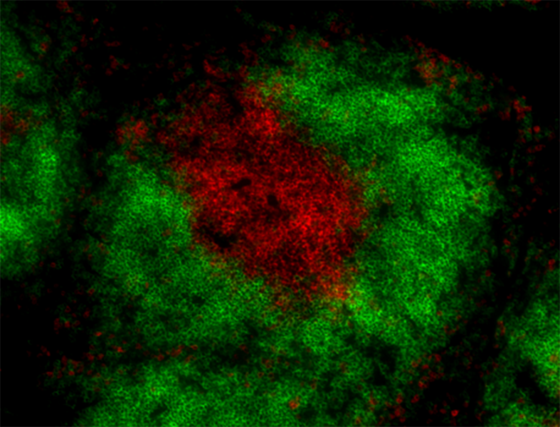
Daudi cells (negative control) (panel A) and HeLa cells (positive control) (panel B) were fixed with 4% paraformaldehyde for 10 minutes, permeabilized with MeOH for 10 minutes, and blocked with 5% FBS for 60 minutes. Cells were then intracellularly stained with 5.0 µg/mL purified anti-YAP1 (clone W19260A) overnight at 4°C, followed by incubation with Alexa Fluor® 594 goat anti-rat IgG (Cat. No. 405422) at 2.0 µg/mL. Nuclei were counterstained with DAPI and the image was captured with a 60X objective. Scale: 50 µm -
IHC staining of purified anti-YAP1 (clone W19260A) on human breast tissue. Tissue was fixed with Fixation Buffer (Cat. No. 420801), blocked with 5% FBS + 5% Normal Serum Block (Cat. No. 927503) and incubated with 10.0 µg/mL purified rat IgG2b, κ isotype control (Cat. No. 400602) (panel A) or 10.0 µg/mL of the primary antibody (panel B) overnight at 4°C. This was followed by incubation with Alexa Fluor® 647 goat anti-rat IgG (Cat. No. 405416) at 2.5 µg/mL. Nuclei were counterstained with DAPI (blue) and the slide was mounted with ProLong™ Gold Antifade Mountant. The image was captured with a 40X objective. Scale: 50 µm -

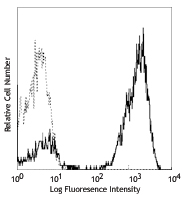
HeLa cells (positive target) were stained with 0.25 µg/test of purified anti-YAP1 (clone W19260A) (filled histogram) or purified rat IgG2b, κ isotype control (Cat. No. 400601) (open histogram) followed by PE goat anti-rat IgG (Cat. No. 405406). -

Daudi cells (negative target) were stained with 0.25 µg/test of purified anti-YAP1 (clone W19260A) (filled histogram) or purified rat IgG2b, κ isotype control (Cat. No. 400601) (open histogram) followed by PE goat anti-rat IgG (Cat. No. 405406).

| Cat # | Size | Price | Quantity Check Availability | Save | ||
|---|---|---|---|---|---|---|
| 603651 | 25 µg | $118 | ||||
| 603652 | 100 µg | $293 | ||||
Yes-associated protein 1, or YAP1, is a transcriptional co-activator vital to cell proliferation and the regulation of apoptosis. YAP1 does not contain a DNA-binding domain and requires interaction with transcriptional factors, particularly TEA domain (TEAD) proteins, to regulate transcription. YAP1 is continuously shuttled between the nucleus, where it is primarily located when Hippo signaling is inactive, and the cytoplasm, where it is sequestered following phosphorylation of its various serine residues by the LATS1/2-MOB1 complex of the Hippo pathway or by kinases AKT, PRP4K, and CDK1. As a key effector of the Hippo signaling pathway along with its paralog TAZ, YAP1 is essential to the regulation of cell growth, stem cell maintenance, organ size, tissue homeostasis, regeneration, embryonic development, and the preservation of genome integrity following DNA damage. YAP1 also functions in the modulation of aerobic glycolysis and glutaminolysis pathways as well as amino acid metabolism. YAP1 can act as a tumor suppressor, usually through its interaction with p53 family proteins and promotion of apoptosis, but most commonly functions as an oncogene. Its overexpression and deregulation are characteristic of several cancers and it has been implicated in the initiation, proliferation, metastasis, metabolic reprogramming, immune escape, and therapy resistance of cancer cells. As such, YAP1 is a potential therapeutic target and prognostic biomarker.
Product DetailsProduct Details
- Verified Reactivity
- Human, Mouse
- Antibody Type
- Monoclonal
- Host Species
- Rat
- Immunogen
- Recombinant human YAP1
- Formulation
- Phosphate-buffered solution, pH 7.2, containing 0.09% sodium azide
- Preparation
- The antibody was purified by affinity chromatography.
- Concentration
- 0.5 mg/mL
- Storage & Handling
- The antibody solution should be stored undiluted between 2°C and 8°C.
- Application
-
WB - Quality tested
ICC, IHC-P, ICFC - Verified - Recommended Usage
-
Each lot of this antibody is quality control tested by western blotting. For western blotting, the suggested use of this reagent is 0.25 - 1.0 µg/mL. For immunocytochemistry, a concentration range of 1.25 - 5.0 μg/mL is recommended. For immunohistochemistry on formalin-fixed paraffin-embedded tissue sections, a concentration range of 5.0 - 10.0 µg/mL is suggested. For intracellular flow cytometric staining, the suggested use of this reagent is 0.03 - 0.5 µg/test. It is recommended that the reagent be titrated for optimal performance for each application.
- Application Notes
-
For ICC, we recommend 4% PFA fixation followed by permeabilization with ice-cold methanol or 0.5% Triton-X. Fixation and permeabilization with only methanol is not recommended.
- RRID
-
AB_2910483 (BioLegend Cat. No. 603651)
AB_2910483 (BioLegend Cat. No. 603652)
Antigen Details
- Structure
- YAP1 is a 504 amino acid protein with a predicted molecular weight of 54 kD.
- Distribution
-
Ubiquitous/ Primarily nuclear localization at low density and primarily cytoplasmic localization at high density
- Function
- Transcription
- Interaction
- TAZ, TEAD1/2/3/4, LATS1/2, YES, SRC, SMAD, p73, RUNX2, PAX3, ERBB4, CK1, ZO1/2
- Biology Area
- Cancer Biomarkers, DNA Repair/Replication, Stem Cells, Transcription Factors
- Antigen References
-
- Chen X, et al. 2020. Front Physiol. 11:389.
- Isfort I, et al. 2019. Sci Rep. 9:19704.
- Raj N and Bam R. 2019. Front. Cell Dev Biol. 7:159.
- Szulzewsky F, et al. 2021. Dev Biol. 475:205-221.
- Gene ID
- 10413 View all products for this Gene ID
- UniProt
- View information about Yap1 on UniProt.org

Related FAQs
Other Formats
View All Yap1 Reagents Request Custom Conjugation| Description | Clone | Applications |
|---|---|---|
| Purified anti-YAP1 | W19260A | WB,IHC-P,ICC,ICFC |
| PE anti-YAP1 | W19260A | ICFC |
Customers Also Purchased
Compare Data Across All Formats
This data display is provided for general comparisons between formats.
Your actual data may vary due to variations in samples, target cells, instruments and their settings, staining conditions, and other factors.
If you need assistance with selecting the best format contact our expert technical support team.
-
Purified anti-YAP1

Whole cell extracts (15 µg total protein) from HeLa, MCF7 (p... 
Whole cell extracts (15 µg total protein) from HeLa cells (p... 
Daudi cells (negative control) (panel A) and HeLa cells (pos... IHC staining of purified anti-YAP1 (clone W19260A) on human ... 
HeLa cells (positive target) were stained with 0.25 µg/test ... 
Daudi cells (negative target) were stained with 0.25 µg/test... -
PE anti-YAP1

HeLa cells (positive control) (filled histogram) and Jurkat ...


Follow Us