 Login/Register


- Clone
- A15158C (See other available formats)
- Regulatory Status
- RUO
- Other Names
- Signal transducer and activator of transcription 1, Transcription factor ISGF-3 components p91/p84
- Isotype
- Mouse IgG1, κ
- Ave. Rating
- Submit a Review
- Product Citations
- publications

-

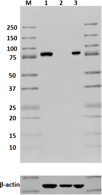
Total cell lysates (15 µg total protein) from U3A (negative control, Lane 1), Jurkat (Lane 2), and HeLa cells treated without (Lane 3) or with (Lane 4) 500 ng/mL nocodazole overnight were resolved by 4-20% Tris-Glycine gel electrophoresis, transferred to nitrocellulose, and probed with 0.25 µg/mL (1:2000 dilution) purified anti-STAT1 clone A15158C (left) antibody and a competitor’s clone used at the manufacturer’s recommended concentration (right). Proteins were visualized by chemiluminescence detection using HRP goat anti-mouse-IgG (Cat. No. 405301) for clone A15158C, and HRP donkey anti-rabbit IgG (Cat. No. 406401) for the competitor’s clone, each at a 1:3000 dilution. Direct-Blot™ HRP anti-β-actin Antibody (Cat. No. 643807) was used as a loading control at a 1:2000 dilution (lower). Lane M: MW ladder. -

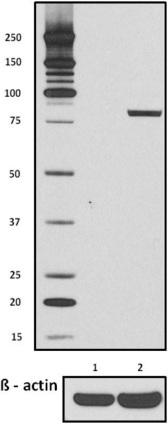
To confirm that the described nocodazole treatment worked, total cell lysates (15 µg total protein) from HeLa cells treated without (-) or with (+) nocodazole 500 ng/mL overnight were resolved by 4-20% Tris-Glycine gel electrophoresis, transferred to nitrocellulose, and probed with 0.5 µg/mL (1:1000 dilution) purified anti-STAT1 Serine 727 antibody (clone A15158B, left) or purified anti-STAT1 antibody (clone A15158C, right). Proteins were visualized by chemiluminescence detection using HRP goat anti-mouse-IgG (Cat. No. 405301). -

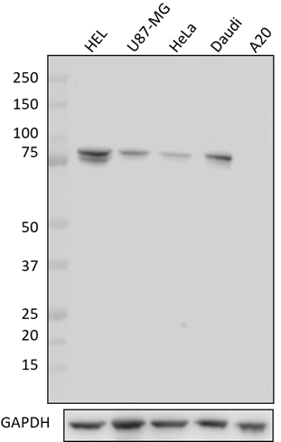
Whole cell extracts (15 µg protein) from the indicated cell lines were resolved by 4-12% Bis-Tris gel electrophoresis, transferred to a PVDF membrane, and probed with 0.05 µg/mL (1:10000 dilution) of Purified anti-STAT1 Antibody, clone A15158C, for 2 hours at room temperature. Proteins were visualized by chemiluminescence detection using HRP goat anti-mouse IgG Antibody (Cat. No. 405306) at a 1:3000 dilution. Direct-Blot™ HRP anti-GAPDH Antibody (Cat. No. 607904) was used as a loading control at a 1:25000 dilution (lower). Lane M: Molecular Weight marker.
| Cat # | Size | Price | Quantity Check Availability | Save | ||
|---|---|---|---|---|---|---|
| 603701 | 25 µg | $101 | ||||
| 603702 | 100 µg | $253 | ||||
STAT1, also known as signal transducter and activator of transcription 1, is a ubiquitously expressed protein that is activated in response to cytokine signaling, including IFN-α, IFN-γ, EGF, PDGF, and IL-6. Upon activation, cytosolic STAT1 is phosphorylated by receptor-associated kinases, translocates to the nucleus, and functions as a transcriptional activator. Two isoforms of STAT1, with predicted molecular weights of 87 (STAT1α) and 83 kD (STAT1β), exist as a result of alternative RNA processing. This clone is specific for STAT1α. STAT1 is involved in IFN-mediated immune responses, and STAT1-deficient mice are highly sensitive to bacterial and viral infections.
Product DetailsProduct Details
- Verified Reactivity
- Human, Mouse
- Antibody Type
- Monoclonal
- Host Species
- Mouse
- Immunogen
- Human STAT1 peptide phosphorylated at Serine 727.
- Formulation
- Phosphate-buffered solution, pH 7.2, containing 0.09% sodium azide.
- Preparation
- The antibody was purified by affinity chromatography.
- Concentration
- 0.5 mg/ml
- Storage & Handling
- The antibody solution should be stored undiluted between 2°C and 8°C.
- Application
-
WB - Quality tested
- Recommended Usage
-
Each lot of this antibody is quality control tested by Western blotting. For Western blotting, the suggested use of this reagent is 0.05 - 0.25 µg per ml (1:2000 - 1:5000 dilution). It is recommended that the reagent be titrated for optimal performance for each application.
- Application Notes
-
This antibody is specific for STAT1 isoform a and is not predicted to recognize the ß isoform due to the absence of the region corresponding to the immunizing peptide.
This clone displayed a stronger affinity for STAT1a in parallel western blot testing with BioLegend’s anti-STAT1 antibody (clone 10C4B40).
U3A cells were selected for antibody specificity testing because the cell line does not express STAT11.
This clone was derived from a human STAT1 peptide phosphorylated at Serine 727 immunogen, but tested negative for phospho-recognition while displaying strong pan-reactivity. Supplemental data is provided confirming the differences in phosphorylation of STAT1 Serine 727 in HeLa cells treated with or without nocodazole. -
Application References
(PubMed link indicates BioLegend citation) -
- Muller M, et al. 1993. EMBO J. 12:4221
- Product Citations
-
- RRID
-
AB_2749867 (BioLegend Cat. No. 603701)
AB_2749868 (BioLegend Cat. No. 603702)
Antigen Details
- Structure
- STAT1α is a 750 amino acid protein with a predicted molecular weight of 87 kD; contains an SH2 domain responsible for homodimerization or heterodimerization.
- Distribution
-
Ubiquitously expressed; cytosolic and nuclear distribution
- Function
- Transcription activator/Cytokine Signaling
- Interaction
- Forms a homodimer or heterodimers with other family members. Interacts with FAK, MCM3, MCM5, TRADD, BRCA1, KIT, IL-27R, IL-2Rβ, IL-2Rγ, IFNαβR, and c-Src.
- Biology Area
- Cell Biology, Signal Transduction, Transcription Factors
- Molecular Family
- Nuclear Markers
- Antigen References
-
- Durbin JE, et al. 1996. Cell 8:443.
- Darnell JE Jr, et al. 1994. Science 264:1415.
- Chen X, et al. 1998. Cell 93:827.
- Ramana CV, et al. 2000. Oncogene 19:2619.
- Gene ID
- 6772 View all products for this Gene ID
- UniProt
- View information about STAT1 on UniProt.org

Related FAQs
Other Formats
View All STAT1 Reagents Request Custom Conjugation| Description | Clone | Applications |
|---|---|---|
| Purified anti-STAT1 | A15158C | WB |
Customers Also Purchased
Compare Data Across All Formats
This data display is provided for general comparisons between formats.
Your actual data may vary due to variations in samples, target cells, instruments and their settings, staining conditions, and other factors.
If you need assistance with selecting the best format contact our expert technical support team.


Follow Us