 Login/Register


- Clone
- 7A6 (See other available formats)
- Regulatory Status
- RUO
- Other Names
- Nuclear factor of activated T-cells, cytoplasmic 1, NFAT transcription complex cytosolic component, NFAT2, NFATc
- Isotype
- Mouse IgG1, κ
- Ave. Rating
- Submit a Review
- Product Citations
- publications

-

Total lysates (15 µg protein) from EL4 (lane 1), HeLa (lane 2), Jurkat (lane 3) and Raw 264.7 cells (lane 4) were resolved by electrophoresis (4-20% Tris-Glycine gel), transferred to nitrocellulose, and probed with 1:500 diluted (1 µg/mL) Purified anti-NFATc1 Antibody, clone 7A6 (upper). Proteins were visualized by chemiluminescence detection using a 1:3000 diluted goat anti-mouse-IgG secondary antibody conjugated to HRP for the anti-NFATc1 Antibody or 1:5000 diluted Direct-Blot HRP anti-β-Actin Antibody, clone 2F1-1(lower). Lane M: Molecular weight ladder. -

Chromatin Immunoprecipitation (ChIP) was performed using Go-ChIP-Grade™ Protein G Enzymatic kit by loading 3 µg of cross-linked chromatin samples from Raw264.7 cells treated with RANKL with either A) 1:500 dilution of Go-ChIP-Grade™ Purified anti-NFATc1 (Clone 7A6), or B) equal amount of Purified Mouse IgG1 Isotype Control Antibody. The enriched DNA was purified and quantified by real-time qPCR using primers targeting mouse TRAP gene region. The amount of immunoprecipitated DNA in each sample is represented as signal relative to the 5% of total amount of input chromatin. -

Chromatin Immunoprecipitation (ChIP) was performed using Go-ChIP-Grade™ Protein G Enzymatic kit by loading 3 µg of cross-linked chromatin samples from Raw264.7 cells treated with RANKL with either A) 1:500 dilution of Go-ChIP-Grade™ Purified anti-NFATc1 (Clone 7A6), or B) equal amount of Purified Mouse IgG1 Isotype Control Antibody. The enriched DNA was purified and quantified by real-time qPCR using primers targeting mouse OSCAR gene region. The amount of immunoprecipitated DNA in each sample is represented as signal relative to the 5% of total amount of input chromatin.
| Cat # | Size | Price | Quantity Check Availability | Save | ||
|---|---|---|---|---|---|---|
| 649601 | 25 µg | $124 | ||||
| 649602 | 100 µg | $268 | ||||
The product of this gene is a component of the nuclear factor of activated T cells DNA-binding transcription complex. The protein complex consists of NFAT1, NFAT2 (NFATc1 or NFATc), NFAT3, and NFAT4. All members of this family are transcription factors with a Rel homology domain and regulate gene transcription in concert with AP-1 (Jun/Fos) to orchestrate an effective immune response. NFAT proteins are predominantly expressed in cells of the immune system but are also expressed in skeletal muscle, keratinocytes and adipocytes, regulating cell differentiation programs in these cells. In resting cells, NFAT proteins are heavily phosphorylated and localized in the cytoplasm. Increased intracellular calcium concentrations activate the calcium/calmodulin-dependent serine phosphatase calcineurin, which dephosphorylates NFAT proteins, resulting in their subsequent translocation to the nucleus.
Proteins belonging to this family of transcription factors play a central role in inducible gene transcription during immune response. The product of this gene is an inducible nuclear component. It functions as a major molecular target for the immunosuppressive drugs such as cyclosporin A. Five transcript variants encoding distinct isoforms have been identified for this gene. Different isoforms of this protein may regulate inducible expression of different cytokine genes.
Product Details
- Verified Reactivity
- Human, Mouse, Rat
- Antibody Type
- Monoclonal
- Host Species
- Mouse
- Immunogen
- Recombinant protein of human NFATc1 amino acids 197-304.
- Formulation
- This antibody is provided in phosphate-buffered solution, pH 7.2, 0.09% sodium azide.
- Preparation
- The antibody was purified by affinity chromatography.
- Concentration
- 0.5 mg/mL
- Storage & Handling
- Upon receipt, store undiluted between 2°C and 8°C.
- Application
-
WB - Quality tested
ChIP - Verified
FC - Reported in the literature, not verified in house
ICC - Reported by the developer, not verified in house - Recommended Usage
-
Each lot of this antibody is quality control tested by Western blotting. For Western blotting applications, a concentration of 1 µg/mL is recommended. For ChIP applications, the suggested dilution is is 1:100-1:2000 by volume. It is recommended that the reagent be titrated for optimal performance for each application.
- Application Notes
-
7A6 antibody detects endogenous human NFATC1 in Western blot. There are 10 isoforms with a predicted MW of 39kD, 74kD, 76kD, 77kD (3), 88kD (2), 100kD, 101kD. With this antibody, observed MW bands range from 70 - 120 kD. The optimal concentration should be determined by titration for each individual assay of interest.
Clone 7A6 Intracellular Staining Procedure:- Prepare the fix/perm solution, consisting of 1% paraformaldehyde in 70% ethanol. Chill for at least one hour at -20°C.
- Suspend cells in cell staining buffer and perform surface stain if necessary.
- Centrifuge for 5 minutes at 1500 RPM and discard supernatant.
- Resuspend cells in 500 µL of fix/perm solution for up to 2 x 10^6 cells and gently vortex.
- Incubate cells on ice for 30-60 minutes in the dark.
- Add 2 mL PBS and centrifuge cells for 1500 RPM for 5 minutes.
- Wash cells 1X with 2 mL cell staining buffer, centrifuge at 1500 RPM for 5 minutes, discard supernatant.
- Adjust cell concentration to about 1 x 10^7 cells/mL, add 100 µL (~ 1 x 10^6 cells) to a FACS tube that already contains the staining antibody and mix gently.
- Incubate 15 minutes at room temperature.
- Wash 2X with cell staining buffer as in step 6.
- Resuspend cells in cell staining buffer and analyze.
-
Application References
(PubMed link indicates BioLegend citation) -
- Timmerman LA, et al. 1997. J. Immunol. 159:2735. (IF)
- Brandt C, et al. 2010. Cytometry A. 77:607. (FC)
- Fan W, et al. 2012. Arthritis Rheum. 64:3715. PubMed
- Product Citations
-
- RRID
-
AB_10680239 (BioLegend Cat. No. 649601)
AB_10679126 (BioLegend Cat. No. 649602)
Antigen Details
- Structure
- 943 amino acids with predicted molecular weight of 101kD. There are 10 isoforms with a predicted MW of 39kD, 74kD, 76kD, 77kD (3), 88kD (2), 100kD, 101kD.
- Distribution
-
Cytoplasm, Nucleus. NFATc1 is cytoplasmic in the phosphorylated form and nuclear after calcineurin-mediated dephosphorylation controlled activation.
- Biology Area
- Cell Biology, Immunology, Neuroscience, Neuroscience Cell Markers, Signal Transduction, Transcription Factors
- Molecular Family
- Nuclear Markers
- Antigen References
-
1. Zhao Q, et al. 2010. Int. J. Biochem. Cell Biol. 42:576.
2. Hoey T, et al. 1995. Immunity 2:461.
3. Northrop JP, et al. 1993. J. Biol. Chem. 268:2917.
4. Hogan PG, et al. 2003. Genes Dev. 17:2205.
5. Shaw KT, et al. 1995. P. Natl. Acad. Sci. USA 92:11205.
6. Serfling E, et al. 2007. Sci. STKE 138:pe42. - Gene ID
- 4772 View all products for this Gene ID
- UniProt
- View information about NFATc1 on UniProt.org

Related FAQs
Other Formats
View All NFATc1 Reagents Request Custom Conjugation| Description | Clone | Applications |
|---|---|---|
| PE anti-NFATc1 | 7A6 | ICFC |
| Purified anti-NFATc1 | 7A6 | WB,FC,ICC,ChIP |
| Alexa Fluor® 488 anti-NFATc1 | 7A6 | ICFC |
Customers Also Purchased
Compare Data Across All Formats
This data display is provided for general comparisons between formats.
Your actual data may vary due to variations in samples, target cells, instruments and their settings, staining conditions, and other factors.
If you need assistance with selecting the best format contact our expert technical support team.
-
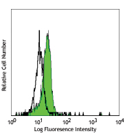
PE anti-NFATc1

Human peripheral blood lymphocytes were fixed and permeabili... -
Purified anti-NFATc1

Total lysates (15 µg protein) from EL4 (lane 1), HeLa (lane ... 
Chromatin Immunoprecipitation (ChIP) was performed using Go-... 
Chromatin Immunoprecipitation (ChIP) was performed using Go-... -
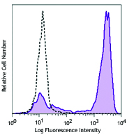
Alexa Fluor® 488 anti-NFATc1

Human peripheral blood lymphocytes were treated with pre-chi...


Follow Us