 Login/Register


- Clone
- BL13756 (See other available formats)
- Regulatory Status
- RUO
- Other Names
- Macrosialin
- Isotype
- Mouse IgG1, κ
- Ave. Rating
- Submit a Review
- Product Citations
- publications

-

Human paraffin-embedded tonsil tissue slices were prepared with a standard protocol of deparaffination and rehydration. Antigen retrieval was done with Citrate buffer 1X pH 6.0 at 95°C for 40 minutes. Tissue was washed with PBS/ 0.05% Tween20 twice for five minutes, blocked with 5% FBS and 0.2% gelatin for 30 minutes and permeabilized with 0.5% Triton X-100. Then, the tissue was stained with 10 µg/mL of purified anti-human CD68 (clone BL13756) at 4°C overnight. On the next day, the tissue was washed twice with PBS and stained with goat anti-mouse secondary antibody (Poly4053) Alexa Fluor™ 594 (red) for two hours at room temperature. The nuclei were counter staining with DAPI (blue). The image was scanned with a 10X objective and stitched with software.
| Cat # | Size | Price | Quantity Check Availability | Save | ||
|---|---|---|---|---|---|---|
| 375602 | 100 µg | $112 | ||||
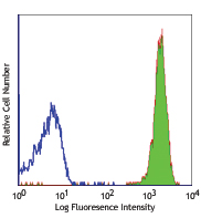
CD68 is a 110 kD glycoprotein, also known as macrosialin, belonging to the sialomucin family. It is closely related to the family of acidic, highly glycosylated lysosomal-associated membrane proteins (LAMPs). CD68 is predominately expressed in cytoplasmic granules of monocytes/macrophages, dendritic cells, and granulocytes. It is one of the useful myeloid cell markers. Further studies have shown that CD68 is also expressed by a subset of hematopoietic progenitors, γ/δ T cells, NK cells, LAK cells, subset of B cells, fibroblasts, and endothelial cells. The biological function of CD68 is still unknown.
Product DetailsProduct Details
- Verified Reactivity
- Human
- Antibody Type
- Monoclonal
- Host Species
- Mouse
- Immunogen
- Synthetic peptide corresponding to residues from the internal region of the human CD68 protein
- Formulation
- Phosphate-buffered solution, pH 7.2, containing 0.09% sodium azide
- Preparation
- The antibody was purified by affinity chromatography
- Concentration
- 0.5 mg/mL
- Storage & Handling
- The antibody solution should be stored undiluted between 2°C and 8°C.
- Application
-
IHC-P - Quality tested
SB - Community verified - Recommended Usage
-
Each lot of this antibody is quality control tested by formalin-fixed paraffin-embedded immunohistochemical staining. For immunohistochemistry, a concentration range of 5 - 10 µg/mL is suggested. It is recommended that the reagent be titrated for optimal performance for each application.
- Additional Product Notes
-
This product has been verified for IHC-P (Immunohistochemistry - formalin-fixed paraffin-embedded tissues) on the NanoString GeoMx® Digital Spatial Profiler. The GeoMx® enables researchers to perform spatial analysis of protein and RNA targets in FFPE and fresh frozen human and mouse samples. For more information about our spatial biology products and the GeoMx® platform, please visit our spatial biology page.
- RRID
-
AB_2876705 (BioLegend Cat. No. 375602)
Antigen Details
- Structure
- Sialomucin family, 110 kD
- Distribution
-
Monocytes/macrophages, dendritic cells, granulocytes, subset of hematopoietic progenitors, γ/δ T cells, NK cells, LAK cells, subset of B cells, fibroblasts, endothelial cells
- Molecular Family
- CD Molecules
- Antigen References
-
1. Holness CL and Simmons DL. 1993. Blood. 81:1607.
2. Gottfried E, et al. 2008. Scand. J. Immunol. 67:453.
3. Hameed A, et al. 1994. Hum. Pathol. 25:872. - Gene ID
- 968 View all products for this Gene ID
- UniProt
- View information about CD68 on UniProt.org


Related FAQs
- If an antibody clone has been previously successfully used in IBEX in one fluorescent format, will other antibody formats work as well?
-
It’s likely that other fluorophore conjugates to the same antibody clone will also be compatible with IBEX using the same sample fixation procedure. Ultimately a directly conjugated antibody’s utility in fluorescent imaging and IBEX may be specific to the sample and microscope being used in the experiment. Some antibody clone conjugates may perform better than others due to performance differences in non-specific binding, fluorophore brightness, and other biochemical properties unique to that conjugate.
- Will antibodies my lab is already using for fluorescent or chromogenic IHC work in IBEX?
-
Fundamentally, IBEX as a technique that works much in the same way as single antibody panels or single marker IF/IHC. If you’re already successfully using an antibody clone on a sample of interest, it is likely that clone will have utility in IBEX. It is expected some optimization and testing of different antibody fluorophore conjugates will be required to find a suitable format; however, legacy microscopy techniques like chromogenic IHC on fixed or frozen tissue is an excellent place to start looking for useful antibodies.
- Are other fluorophores compatible with IBEX?
-
Over 18 fluorescent formats have been screened for use in IBEX, however, it is likely that other fluorophores are able to be rapidly bleached in IBEX. If a fluorophore format is already suitable for your imaging platform it can be tested for compatibility in IBEX.
- The same antibody works in one tissue type but not another. What is happening?
-
Differences in tissue properties may impact both the ability of an antibody to bind its target specifically and impact the ability of a specific fluorophore conjugate to overcome the background fluorescent signal in a given tissue. Secondary stains, as well as testing multiple fluorescent conjugates of the same clone, may help to troubleshoot challenging targets or tissues. Using a reference control tissue may also give confidence in the specificity of your staining.
- How can I be sure the staining I’m seeing in my tissue is real?
-
In general, best practices for validating an antibody in traditional chromogenic or fluorescent IHC are applicable to IBEX. Please reference the Nature Methods review on antibody based multiplexed imaging for resources on validating antibodies for IBEX.
Other Formats
View All Reagents Request Custom Conjugation| Description | Clone | Applications |
|---|---|---|
| Purified anti-human CD68 | BL13756 | IHC-P,SB |
| TotalSeq™-Bn1304 anti-human CD68 | BL13756 | SB |
| Alexa Fluor® 647 anti-human CD68 | BL13756 | IHC-P |
Customers Also Purchased

Compare Data Across All Formats
This data display is provided for general comparisons between formats.
Your actual data may vary due to variations in samples, target cells, instruments and their settings, staining conditions, and other factors.
If you need assistance with selecting the best format contact our expert technical support team.
-
Purified anti-human CD68

Human paraffin-embedded tonsil tissue slices were prepared w... -
TotalSeq™-Bn1304 anti-human CD68
-
Alexa Fluor® 647 anti-human CD68

Human paraffin-embedded prostate tissue slices were prepared... 
Human paraffin-embedded tonsil tissue slices were prepared w...


Follow Us