 Login/Register


- Clone
- 6/40c (See other available formats)
- Regulatory Status
- RUO
- Other Names
- CEACAM8, CD67, CGM6, NCA-95
- Isotype
- Mouse IgG1, κ
- Ave. Rating
- Submit a Review
- Product Citations
- publications

-

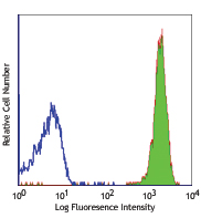
Human peripheral blood granulocytes was stained with CD66b (clone 6/40c) purified (filled histogram) or mouse IgG1, κ isotype control (clone MOPC-21) purified (open histogram) followed by Poly4053 PE. -

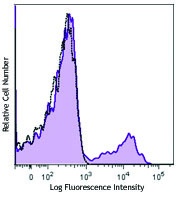
Human paraffin-embedded spleen tissue slices were prepared with a standard protocol of deparaffinization and rehydration. Antigen retrieval was done with Sodium Citrate H.I.E.R., 1x pH 6.0 at 95°C for 40 minutes. Tissue was washed with PBS/0.05% Tween 20 twice for five minutes and blocked with 5% FBS and 0.2% gelatin for 30 minutes. Then, the tissue was stained with 10 µg/ml of purified anti-human CD66b (clone 6/40c) antibody overnight at 4°C. On the next day, tissue was incubate with Alexa Fluor® 594 Goat anti-mouse IgG (clone poly4053) antibody (red). Nuclei were counter-stained with DAPI (blue). The image was scanned with a 10X objective and stitched with MetaMorph® software. -

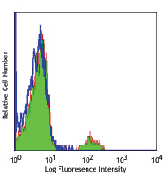
SeqIF™ (sequential immunofluorescence) staining on COMET™ of Purified anti-CD66b (clone 6/40c, yellow) on formalin-fixed paraffin-embedded human tonsil tissue at 5 µg/mL. Alexa Fluor™ Plus 647 Goat anti-Mouse IgG antibody (Lunaphore, Cat. No. DR647MS) was used as a secondary antibody. Nuclei were counterstained with DAPI (blue). Tissue underwent an all-in-one dewaxing and antigen retrieval preprocessing.
| Cat # | Size | Price | Quantity Check Availability | Save | ||
|---|---|---|---|---|---|---|
| 392902 | 100 µg | $141 | ||||
CD66b is a 95-100 kD glycosylphosphatidylinositol (GPI)-linked protein also known as CD67, CGM6, and NCA-95. CD66b is a member of the immunoglobulin superfamily, carcinoembryonic antigen (CEA)-like subfamily. CD66b, expressed on granulocytes, has been reported to induce activation in neutrophils and to be involved in heterophilic adhesion with CD66c.
Product DetailsProduct Details
- Verified Reactivity
- Human
- Antibody Type
- Monoclonal
- Host Species
- Mouse
- Immunogen
- Recombinant soluble human CEACAM8-Fc produced in HEK293 cells
- Formulation
- Phosphate-buffered solution, pH 7.2, containing 0.09% sodium azide.
- Preparation
- The antibody was purified by affinity chromatography.
- Concentration
- 0.5 mg/ml
- Storage & Handling
- The antibody solution should be stored undiluted between 2°C and 8°C.
- Application
-
FC - Quality tested
IHC-P - Verified
SB - Community verified - Recommended Usage
-
Each lot of this antibody is quality control tested by immunofluorescent staining with flow cytometric analysis. For flow cytometric staining, the suggested use of this reagent is ≤ 0.5 µg per million cells in 100 µl volume. For immunohistochemistry, a concentration range of 5.0 - 10 µg/ml is suggested. It is recommended that the reagent be titrated for optimal performance for each application.
- Additional Product Notes
-
For the use of this antibody in spatial biology applications, we have partnered with Lunaphore Technologies for demonstration of our antibodies on the COMET™. The COMET™ platform is an automated, end-to-end spatial biology solution developed for rapid and flexible multiplex tissue profiling. More information on the COMET™ and a complete list of our antibodies that have been demonstrated on the COMET™ can be found here.
-
Application References
(PubMed link indicates BioLegend citation) -
- Singer, et al. 2014. PLoS One. 9(4):e94106. PMID: 24743304
- Klaile, et al. 2013. Respir Res. 14:85. PMID 23941132
- Product Citations
-
- RRID
-
AB_2728422 (BioLegend Cat. No. 392902)
Antigen Details
- Structure
- Ig superfamily, CEA antigen group, GPI-linked glycoprotein
- Distribution
-
Granulocytes
- Function
- Cell adhesion, neutrophil activation
- Ligand/Receptor
- CD66c
- Cell Type
- Granulocytes
- Biology Area
- Immunology
- Molecular Family
- Adhesion Molecules, CD Molecules
- Antigen References
-
- Kuijpers T, et al. 1993. J Immunol. 151:4934.
- Kuroki M, et al. 1992. J Leuk Biol. 52:551.
- Gene ID
- 1088 View all products for this Gene ID
- UniProt
- View information about CD66b on UniProt.org


Related FAQs
- If an antibody clone has been previously successfully used in IBEX in one fluorescent format, will other antibody formats work as well?
-
It’s likely that other fluorophore conjugates to the same antibody clone will also be compatible with IBEX using the same sample fixation procedure. Ultimately a directly conjugated antibody’s utility in fluorescent imaging and IBEX may be specific to the sample and microscope being used in the experiment. Some antibody clone conjugates may perform better than others due to performance differences in non-specific binding, fluorophore brightness, and other biochemical properties unique to that conjugate.
- Will antibodies my lab is already using for fluorescent or chromogenic IHC work in IBEX?
-
Fundamentally, IBEX as a technique that works much in the same way as single antibody panels or single marker IF/IHC. If you’re already successfully using an antibody clone on a sample of interest, it is likely that clone will have utility in IBEX. It is expected some optimization and testing of different antibody fluorophore conjugates will be required to find a suitable format; however, legacy microscopy techniques like chromogenic IHC on fixed or frozen tissue is an excellent place to start looking for useful antibodies.
- Are other fluorophores compatible with IBEX?
-
Over 18 fluorescent formats have been screened for use in IBEX, however, it is likely that other fluorophores are able to be rapidly bleached in IBEX. If a fluorophore format is already suitable for your imaging platform it can be tested for compatibility in IBEX.
- The same antibody works in one tissue type but not another. What is happening?
-
Differences in tissue properties may impact both the ability of an antibody to bind its target specifically and impact the ability of a specific fluorophore conjugate to overcome the background fluorescent signal in a given tissue. Secondary stains, as well as testing multiple fluorescent conjugates of the same clone, may help to troubleshoot challenging targets or tissues. Using a reference control tissue may also give confidence in the specificity of your staining.
- How can I be sure the staining I’m seeing in my tissue is real?
-
In general, best practices for validating an antibody in traditional chromogenic or fluorescent IHC are applicable to IBEX. Please reference the Nature Methods review on antibody based multiplexed imaging for resources on validating antibodies for IBEX.
Other Formats
View All CD66b Reagents Request Custom Conjugation| Description | Clone | Applications |
|---|---|---|
| Purified anti-human CD66b | 6/40c | FC,IHC-P,SB |
| PE anti-human CD66b | 6/40c | FC |
| TotalSeq™-A0166 anti-human CD66b | 6/40c | PG |
| Alexa Fluor® 594 anti-human CD66b | 6/40c | IHC-P |
| TotalSeq™-C0166 anti-human CD66b | 6/40c | PG |
| Alexa Fluor® 647 anti-human CD66b | 6/40c | IHC-P,FC |
| TotalSeq™-B0166 anti-human CD66b | 6/40c | PG |
| PE/Fire™ 640 anti-human CD66b Antibody | 6/40c | FC |
| Brilliant Violet 421™ anti-human CD66b Antibody | 6/40c | FC,IHC-P |
| TotalSeq™-D0166 anti-human CD66b | 6/40c | PG |
| TotalSeq™-Bn0166 anti-human CD66b | 6/40c | SB |
Customers Also Purchased
Compare Data Across All Formats
This data display is provided for general comparisons between formats.
Your actual data may vary due to variations in samples, target cells, instruments and their settings, staining conditions, and other factors.
If you need assistance with selecting the best format contact our expert technical support team.
-
Purified anti-human CD66b

Human peripheral blood granulocytes was stained with CD66b (... 
Human paraffin-embedded spleen tissue slices were prepared w... 
SeqIF™ (sequential immunofluorescence) staining on COMET™ of... -
PE anti-human CD66b

Human peripheral blood granulocytes were stained with CD66b ... -
TotalSeq™-A0166 anti-human CD66b
-
Alexa Fluor® 594 anti-human CD66b

Human paraffin-embedded spleen tissue slices were prepared w... -
TotalSeq™-C0166 anti-human CD66b
-
Alexa Fluor® 647 anti-human CD66b

Human paraffin-embedded spleen tissue slices were prepared w... 
Human peripheral blood granulocytes were stained with CD66b ... -
TotalSeq™-B0166 anti-human CD66b
-
PE/Fire™ 640 anti-human CD66b Antibody

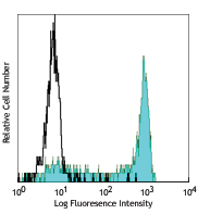
Human peripheral blood granulocytes were stained with anti-h... -
Brilliant Violet 421™ anti-human CD66b Antibody

Human peripheral blood granulocytes were stained with CD66b ... 
Human paraffin-embedded spleen tissue slices were prepared w... -
TotalSeq™-D0166 anti-human CD66b
-
TotalSeq™-Bn0166 anti-human CD66b


Follow Us