 Login/Register


- Clone
- OKT3 (See other available formats)
- Regulatory Status
- RUO
- Workshop
- HCDM listed
- Other Names
- T3, CD3ε
- Isotype
- Mouse IgG2a, κ
- Ave. Rating
- Submit a Review
- Product Citations
- publications

-

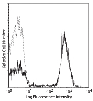
Human peripheral blood lymphocytes stained with purified OKT3, followed by anti-mouse IgGs-FITC
| Cat # | Size | Price | Quantity Check Availability | Save | ||
|---|---|---|---|---|---|---|
| 317301 | 25 µg | $30 | ||||
| 317302 | 100 µg | $55 | ||||
CD3ε is a 20 kD chain of the CD3/T cell receptor (TCR) complex, which is composed of two CD3ε, one CD3γ, one CD3δ, one CD3ζ (CD247), and a T cell receptor (α/β or γ/δ) heterodimer. It is found on all mature T lymphocytes, NK T cells, and some thymocytes. CD3, also known as T3, is a member of the immunoglobulin superfamily that plays a role in antigen recognition, signal transduction, and T cell activation.
Product DetailsProduct Details
- Verified Reactivity
- Human
- Antibody Type
- Monoclonal
- Host Species
- Mouse
- Formulation
- Phosphate-buffered solution, pH 7.2, containing 0.09% sodium azide.
- Preparation
- The antibody was purified by affinity chromatography.
- Concentration
- 0.5 mg/ml
- Storage & Handling
- The antibody solution should be stored undiluted between 2°C and 8°C, and protected from prolonged exposure to light. Do not freeze.
- Application
-
FC - Quality tested
IHC-F - Reported in the literature, not verified in house - Recommended Usage
-
Each lot of this antibody is quality control tested by immunofluorescent staining with flow cytometric analysis. For flow cytometric staining, the suggested use of this reagent is ≤0.25 µg per million cells in 100 µl volume. It is recommended that the reagent be titrated for optimal performance for each application.
- Application Notes
-
The OKT3 monoclonal antibody reacts with an epitope on the epsilon-subunit within the human CD3 complex.
Clone OKT3 can block the binding of clones SK7 and UCHT1.4 The OKT3 antibody is able to induce T cell activation. Additional reported applications (for the relevant formats) include: immunohistochemical staining of acetone-fixed frozen sections and activation of T cells. The LEAF™ purified antibody (Endotoxin <0.1 EU/µg, Azide-Free, 0.2 µm filtered) is recommended for functional assays (Cat. No. 317304). For highly sensitive assays, we recommend Ultra-LEAF™ purified antibody (Cat. No. 317326) with a lower endotoxin limit than standard LEAF™ purified antibodies (Endotoxin <0.01 EU/µg). -
Application References
(PubMed link indicates BioLegend citation) -
- Schlossman S, et al. Eds. 1995. Leucocyte Typing V. Oxford University Press. New York.
- Knapp W. 1989. Leucocyte Typing IV. Oxford University Press New York.
- Barclay N, et al. 1997. The Leucocyte Antigen Facts Book. Academic Press Inc. San Diego.
- Li B, et al. 2005. Immunology 116:487.
- Jeong HY, et al. 2008. J. Leuckocyte Biol. 83:755. PubMed
- Alter G, et al. 2008. J. Virol. 82:9668. PubMed
- Manevich-Mendelson E, et al. 2009. Blood 114:2344. PubMed
- Pinto JP, et al. 2010. Immunology. 130:217. PubMed
- Biggs MJ, et al. 2011. J. R. Soc. Interface. 8:1462. PubMed
- Product Citations
-
- RRID
-
AB_571926 (BioLegend Cat. No. 317301)
AB_571927 (BioLegend Cat. No. 317302)
Antigen Details
- Structure
- Ig superfamily, the subunits CD3γ, CD3δ, CD3ζ (CD247) and TCR (α/β or γ/δ) form the CD3/TCR complex, 20 kD
- Distribution
-
Mature T and NK T cells, thymocyte differentiation
- Function
- Antigen recognition, signal transduction, T cell activation
- Ligand/Receptor
- Peptide antigen bound to MHC
- Cell Type
- NKT cells, T cells, Thymocytes, Tregs
- Biology Area
- Immunology
- Molecular Family
- CD Molecules
- Antigen References
-
- Barclay N, et al. 1993. The Leucocyte FactsBook. Academic Press. San Diego.
- Beverly P, et al. 1981. Eur. J. Immunol. 11:329.
- Lanier L, et al. 1986. J. Immunol. 137:2501.
- Gene ID
- 916 View all products for this Gene ID
- UniProt
- View information about CD3 on UniProt.org


Other Formats
View All CD3 Reagents Request Custom ConjugationCustomers Also Purchased
Compare Data Across All Formats
This data display is provided for general comparisons between formats.
Your actual data may vary due to variations in samples, target cells, instruments and their settings, staining conditions, and other factors.
If you need assistance with selecting the best format contact our expert technical support team.
-
Purified anti-human CD3

Human peripheral blood lymphocytes stained with purified OKT... -
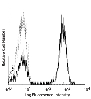
FITC anti-human CD3

Human peripheral blood lymphocytes stained with OKT3 FITC -
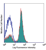
PE anti-human CD3

Human peripheral blood lymphocytes stained with OKT3 PE 
Human peripheral blood was stained with CD3 (clone OKT3) PE ... -
Alexa Fluor® 488 anti-human CD3

Human peripheral blood lymphocytes stained with OKT3 Alexa F... -
Alexa Fluor® 647 anti-human CD3

Human peripheral blood lymphocytes stained with OKT3 Alexa F... -
Pacific Blue™ anti-human CD3

Human peripheral blood lymphocytes were stained with CD3 (cl... -
APC anti-human CD3

Human peripheral blood lymphocytes stained with OKT3 APC -
Biotin anti-human CD3

Human peripheral lymphocytes were stained with biotinylated ... -
Brilliant Violet 605™ anti-human CD3

Human peripheral lymphocytes were stained with CD3 (clone OK... -
Brilliant Violet 650™ anti-human CD3

Human peripheral lymphocytes were stained with CD3 (clone OK... -
Ultra-LEAF™ Purified anti-human CD3

Human peripheral blood lymphocytes stained with LEAF™ purifi... -
Brilliant Violet 711™ anti-human CD3

Human peripheral lymphocytes were stained with CD3 (clone OK... -
Brilliant Violet 785™ anti-human CD3

Human peripheral lymphocytes were stained with CD3 (clone OK... -
Brilliant Violet 510™ anti-human CD3

Human peripheral blood lymphocytes were stained with CD3 (cl... -
PE/Cyanine7 anti-human CD3

Human peripheral blood lymphocytes were stained with CD3 (cl... -
PerCP/Cyanine5.5 anti-human CD3

Human peripheral blood lymphocytes were stained with CD3 (cl... -
PerCP anti-human CD3

Human peripheral blood lymphocytes were stained with CD3 (cl... -
Alexa Fluor® 700 anti-human CD3

Human peripheral blood lymphocytes were stained with CD3 (cl... -
APC/Cyanine7 anti-human CD3

Human peripheral blood lymphocytes were stained with CD20 FI... -
Brilliant Violet 421™ anti-human CD3

Human peripheral blood lymphocytes were stained with CD3 (cl... -
PE/Dazzle™ 594 anti-human CD3

Human peripheral blood lymphocytes were stained with CD3 (cl... -
APC/Fire™ 750 anti-human CD3

Human peripheral blood lymphocytes were stained with CD20 FI... -
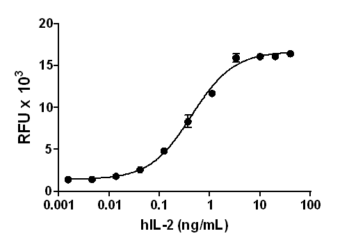
GMP Ultra-LEAF™ Purified anti-human CD3 SF

PBMC derived T cells were activated in the presence of Ulltr... -
PE/Cyanine5 anti-human CD3 Antibody

Human peripheral blood lymphocytes were stained with CD20 (c... -
GMP Ultra-LEAF™ Biotin anti-human CD3 SF

Human peripheral lymphocytes were stained with GMP Ultra-LEA... 
PBMC-derived T cells were activated in the presence of GMP U... -
Spark UV™ 387 anti-human CD3

Human peripheral blood lymphocytes were stained with anti-hu... -
Spark Red™ 718 anti-human CD3

Human peripheral blood lymphocytes were stained with anti-hu... -
TotalSeq™-A1289 anti-human CD3
-
TotalSeq™-B1289 anti-human CD3
-
TotalSeq™-C1289 anti-human CD3


Follow Us