 Login/Register


- Clone
- OX-8 (See other available formats)
- Regulatory Status
- RUO
- Other Names
- CD8a, T8, Leu-8, Ly-2, Lyt2
- Isotype
- Mouse IgG1, κ
- Ave. Rating
- Submit a Review
- Product Citations
- publications

-

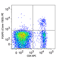
LOU rat splenocytes were stained with CD3 (clone 1F4) APC and CD8a (clone OX-8) PE/Cyanine7 (left) or Mouse IgG1, ? PE/Cyanine7 isotype control (right).
| Cat # | Size | Price | Quantity Check Availability | Save | ||
|---|---|---|---|---|---|---|
| 201715 | 25 µg | $118 | ||||
| 201716 | 100 µg | $276 | ||||
CD8a is a 32 kD glycoprotein also known as T8, Lyt2, Ly-2, and CD8α. CD8a is a member of the immunoglobulin superfamily expressed on most thymocytes, subset of mature T cells, most NK cells, macrophages, and some activated CD4+ T cells (not resting). CD8a forms heterodimers with the CD8β chain (CD8b) on the surface of most thymocytes, while mature peripheral T lymphocytes express almost exclusively the CD8 αβ heterodimer. Intestinal intraepithelial lymphocytes express CD8a without CD8b. CD8 is an antigen co-receptor on T cells that interacts with MHC class I on antigen-presenting cells or epithelial cells. CD8 participates in T cell activation through its association with the T cell receptor complex and protein tyrosine kinase lck (p56lck).
Product DetailsProduct Details
- Verified Reactivity
- Rat
- Antibody Type
- Monoclonal
- Host Species
- Mouse
- Immunogen
- High molecular weight glycoproteins from rat thymocytes
- Formulation
- Phosphate-buffered solution, pH 7.2, containing 0.09% sodium azide.
- Preparation
- The antibody was purified by affinity chromatography and conjugated with PE/Cyanine7 under optimal conditions.
- Concentration
- 0.2 mg/ml
- Storage & Handling
- The antibody solution should be stored undiluted between 2°C and 8°C, and protected from prolonged exposure to light. Do not freeze.
- Application
-
FC - Quality tested
- Recommended Usage
-
Each lot of this antibody is quality control tested by immunofluorescent staining with flow cytometric analysis. For flow cytometric staining, the suggested use of this reagent is =0.25 µg per million cells in 100 µl volume. It is recommended that the reagent be titrated for optimal performance for each application.
- Excitation Laser
-
Blue Laser (488 nm)
Green Laser (532 nm)/Yellow-Green Laser (561 nm)
- Application Notes
-
The OX-8 antibody has been reported to partially block T cell responses, including mixed lymphocyte reactions and cytotoxic T cell responses, and to induce macrophage activation. Additional reported applications (for the relevant formats) include: immunohistochemistry1,2 of acetone-fixed frozen sections and formalin-fixed paraffin-embedded sections, immunoprecipitation3, in vivo and in vitro blocking of T cell responses3, 4, macrophage stimulation5, and Western blotting3,6.
-
Application References
(PubMed link indicates BioLegend citation) -
- Barclay AN. 1981. Immunology 42:593. (IHC)
- Wallgren AC, et al. 1995. Transplantation 60:594. (IHC)
- Torres-Nagel N, et al. 1992. Eur. J. Immunol. 22:2841. (IP, WB)
- Mason DW, et al. 1983. Immunol. Rev. 74:57.
- Hirji N, et al. 1997. J. Immunol. 158:1833. (FA)
- Mitnacht R,, et al. 1998. J. Immunol. 160:700. (WB)
- RRID
-
AB_2814099 (BioLegend Cat. No. 201715)
AB_2814100 (BioLegend Cat. No. 201716)
Antigen Details
- Structure
- Ig superfamily, approximately 32 kD
- Distribution
-
Thymocytes, NK cells, most CD8+ cells, intestinal intraepithelial lymphocytes, activated CD4+ cells, macrophages
- Function
- T cell activation, co-receptor for T cell activation
- Ligand/Receptor
- MHC class I molecule
- Cell Type
- Lymphocytes, Macrophages, NK cells, Thymocytes
- Biology Area
- Immunology
- Molecular Family
- CD Molecules
- Antigen References
-
1. Johnson P, et al. 1985. EMBO J. 4:2539.
2. Thomas ML, et al. 1983. Eur. J. Immunol. 13:855. - Gene ID
- 24930 View all products for this Gene ID
- UniProt
- View information about CD8alpha on UniProt.org
Related Pages & Pathways
Pathways


Other Formats
View All CD8a Reagents Request Custom Conjugation| Description | Clone | Applications |
|---|---|---|
| Purified anti-rat CD8a | OX-8 | FC,IHC-F,IP,WB |
| FITC anti-rat CD8a | OX-8 | FC |
| PE anti-rat CD8a | OX-8 | FC |
| Alexa Fluor® 647 anti-rat CD8a | OX-8 | FC,IHC-F |
| PerCP anti-rat CD8a | OX-8 | FC |
| PE/Cyanine7 anti-rat CD8a | OX-8 | FC |
Customers Also Purchased
Compare Data Across All Formats
This data display is provided for general comparisons between formats.
Your actual data may vary due to variations in samples, target cells, instruments and their settings, staining conditions, and other factors.
If you need assistance with selecting the best format contact our expert technical support team.
-
Purified anti-rat CD8a
-
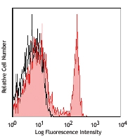
FITC anti-rat CD8a

LOU rat splenocytes stained with OX-8 FITC -
PE anti-rat CD8a

LOU rat splenocytes stained with OX-8 PE -
Alexa Fluor® 647 anti-rat CD8a

LOU rat splenocytes stained with OX-8 Alexa Fluor® 647 
Rat frozen spleen section was fixed with 4% paraformaldehyde... -
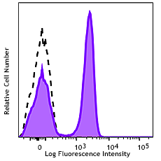
PerCP anti-rat CD8a

LOU rat splenocytes stained with OX-8 PerCP -
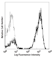
PE/Cyanine7 anti-rat CD8a

LOU rat splenocytes were stained with CD3 (clone 1F4) APC an...


Follow Us