 Login/Register


- Clone
- CD7-6B7 (See other available formats)
- Regulatory Status
- RUO
- Workshop
- IV T-164
- Other Names
- gp40
- Isotype
- Mouse IgG2a, κ
- Ave. Rating
- Submit a Review
- Product Citations
- publications

-

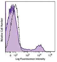
Human peripheral blood lymphocytes were stained with CD7 (clone CD7-6B7) PE/Cyanine7 (filled histogram) or mouse IgG2a, κ PE/Cyanine7 isotype control (open histogram).
| Cat # | Size | Price | Quantity Check Availability | Save | ||
|---|---|---|---|---|---|---|
| 343113 | 25 tests | $112 | ||||
| 343114 | 100 tests | $265 | ||||
CD7 is a 40 kD type I transmembrane glycoprotein also known as gp40. It is a member of the immunoglobulin superfamily found on T cells, NK cells, thymocytes, hematopoietic progenitors, and monocytes (weakly). CD7 is also expressed on acute lymphocytic leukemia (ALL) and some acute myeloid leukemia (AML) cells. CD7 crosslinking induces a calcium flux in T lymphocytes, presumably as a result of cytoplasmic domain association with PI3-kinase. CD7 costimulation can induce cytokine secretion and modulate cellular adhesion.
Product DetailsProduct Details
- Verified Reactivity
- Human
- Antibody Type
- Monoclonal
- Host Species
- Mouse
- Immunogen
- KG1a cell line
- Formulation
- Phosphate-buffered solution, pH 7.2, containing 0.09% sodium azide and BSA (origin USA)
- Preparation
- The antibody was purified by affinity chromatography and conjugated with PE/Cyanine7 under optimal conditions.
- Concentration
- Lot-specific (to obtain lot-specific concentration and expiration, please enter the lot number in our Certificate of Analysis online tool.)
- Storage & Handling
- The antibody solution should be stored undiluted between 2°C and 8°C, and protected from prolonged exposure to light. Do not freeze.
- Application
-
FC - Quality tested
- Recommended Usage
-
Each lot of this antibody is quality control tested by immunofluorescent staining with flow cytometric analysis. For flow cytometric staining, the suggested use of this reagent is 5 µl per million cells in 100 µl staining volume or 5 µl per 100 µl of whole blood.
- Excitation Laser
-
Blue Laser (488 nm)
Green Laser (532 nm)/Yellow-Green Laser (561 nm)
- Application Notes
-
Additional reported (for the relevant formats) applications include proteogenomics2.
- Additional Product Notes
- BioLegend is in the process of converting the name PE/Cy7 to PE/Cyanine7. The dye molecule remains the same, so you should expect the same quality and performance from our PE/Cyanine7 products. Please contact Technical Service if you have any questions.
-
Application References
(PubMed link indicates BioLegend citation) -
- Knapp W, et al. 1989. Leucocyte Typing IV:White Cell Differentiation Antigens. Oxford University Press.
- Peterson VM, et al. 2017. Nat. Biotechnol. 35:936. (PG)
- Product Citations
-
- RRID
-
AB_2563940 (BioLegend Cat. No. 343113)
AB_2563941 (BioLegend Cat. No. 343114)
Antigen Details
- Structure
- Ig superfamily, type I transmembrane glycoprotein, 40 kD
- Distribution
-
T cells, NK cells, thymocytes, hematopoietic progenitors, myeloid leukemic cell subsets
- Function
- T cell activation
- Ligand/Receptor
- Cytoplasmic domain associates with P13-kinase
- Cell Type
- Hematopoietic stem and progenitors, Leukemia, NK cells, T cells, Thymocytes
- Biology Area
- Costimulatory Molecules, Immunology
- Molecular Family
- CD Molecules
- Antigen References
-
1. Barclay N, et al. 1993. The Leucocyte Antigen FactsBook. Academic Press Inc. San Diego.
2. Stillwell R, et al. 2001. Immunol. Res. 24:31.
3. Rabinowich H, et al. 1994. J. Immunol. 152:517. - Gene ID
- 924 View all products for this Gene ID
- UniProt
- View information about CD7 on UniProt.org
Related Pages & Pathways
Pathways


Related FAQs
Other Formats
View All CD7 Reagents Request Custom ConjugationCustomers Also Purchased
Compare Data Across All Formats
This data display is provided for general comparisons between formats.
Your actual data may vary due to variations in samples, target cells, instruments and their settings, staining conditions, and other factors.
If you need assistance with selecting the best format contact our expert technical support team.
-
Purified anti-human CD7
-
FITC anti-human CD7

Human peripheral blood lymphocytes stained with mouse IgG2a,... 
Human peripheral blood lymphocytes stained with CD7-6B7 FITC... -
PE anti-human CD7

Human peripheral blood lymphocytes stained with CD7 (clone C... -
APC anti-human CD7

Human peripheral blood lymphocytes stained with CD7 (clone C... -
PE/Cyanine5 anti-human CD7

Human peripheral blood lymphocytes stained with CD7-6B7 PE/C... -
PE/Cyanine7 anti-human CD7

Human peripheral blood lymphocytes were stained with CD7 (cl... -
Purified anti-human CD7 (Maxpar® Ready)

Human PBMCs stained with 145Nd-anti-CD4 (RPA-T4) and 147Sm-a... -
PerCP/Cyanine5.5 anti-human CD7

Human peripheral blood lymphocytes were stained with CD7 (cl... -
PE/Dazzle™ 594 anti-human CD7

Human peripheral blood lymphocytes were stained with CD7 (cl... -
APC/Fire™ 750 anti-human CD7

Human peripheral blood lymphocytes were stained with CD7 (cl... -
APC anti-human CD7

Typical results from human peripheral blood lymphocytes stai... -
FITC anti-human CD7

Typical results from human peripheral blood lymphocytes stai... -
Alexa Fluor® 647 anti-human CD7

Human peripheral blood lymphocytes were stained with CD7 (cl... -
TotalSeq™-A0066 anti-human CD7
-
Alexa Fluor® 700 anti-human CD7

Human peripheral blood lymphocytes were stained with CD7 (cl... -
TotalSeq™-C0066 anti-human CD7
-
PE anti-human CD7
Typical results from human peripheral blood lymphocytes stai... -
TotalSeq™-B0066 anti-human CD7
-
Pacific Blue™ anti-human CD7 Antibody

Human peripheral blood lymphocytes were stained with anti-hu... -
TotalSeq™-D0066 anti-human CD7
-
GMP APC anti-human CD7

Typical results from human peripheral blood lymphocytes stai... -
GMP FITC anti-human CD7

Typical results from human peripheral blood lymphocytes stai... -
PE/Cyanine7 anti-human CD7

Typical results from human peripheral blood lymphocytes stai... -
Pacific Blue™ anti-human CD7

Typical results from human peripheral blood lymphocytes stai... -
GMP PE anti-human CD7

Typical results from human peripheral blood lymphocytes stai... -
PE/Dazzle™ 594 anti-human CD7

Typical results from human peripheral blood lymphocytes stai... -
GMP PE/Cyanine7 anti-human CD7

Typical results from human peripheral blood lymphocytes stai... -
GMP Pacific Blue™ anti-human CD7

Typical results from human peripheral blood lymphocytes stai... -
Spark NIR™ 685 anti-human CD7

Human peripheral blood lymphocytes were stained with anti-hu...


Follow Us